

Abnormal expression of key genes and proteins in the canonical Wnt/β-catenin pathway of articular cartilage in a rat model of exercise-induced osteoarthritis
- PMID: 26794964
- PMCID: PMC4768959
- DOI: 10.3892/mmr.2016.4798
Abnormal expression of key genes and proteins in the canonical Wnt/β-catenin pathway of articular cartilage in a rat model of exercise-induced osteoarthritis
Abstract
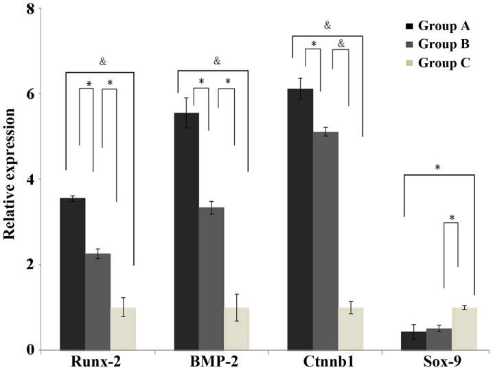
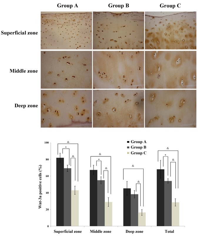
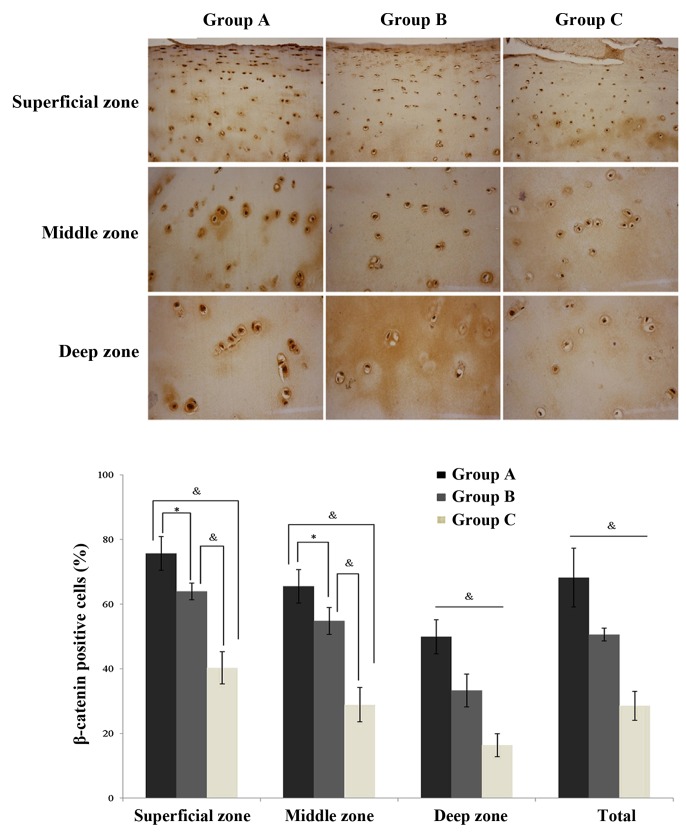
To investigate the molecular pathogenesis of the canonical Wnt/β-catenin pathway in exercise-induced osteoarthritis (OA), 30 male healthy Sprague Dawley rats were divided into three groups (control, normal exercise‑induced OA and injured exercise‑induced OA groups) in order to establish the exercise‑induced OA rat model. The mRNA and protein expression levels of Runx‑2, BMP‑2, Ctnnb1, Sox‑9, collagen Ⅱ, Mmp‑13, Wnt‑3a and β‑catenin in chondrocytes were detected by reverse transcription‑quantitative polymerase chain reaction, western blotting and immunohistochemical staining. The mRNA levels of Runx‑2, BMP‑2 and Ctnnb1 were upregulated in the normal exercise‑induced OA and injured exercise‑induced OA groups; while Runx‑2 and BMP‑2 were upregulated in the injured exercise‑induced OA group when compared with the normal exercise‑induced OA group. The protein levels of Mmp‑13, Wnt‑3a and β‑catenin were increased and collagen Ⅱ was reduced in the normal exercise‑induced OA and injured exercise‑induced OA groups. Ctnnb1, Wnt‑3a and β‑catenin, which are key genes and proteins in the canonical Wnt/β‑catenin pathway, were abnormally expressed in chondrocytes of the exercise‑induced OA rat model. Ctnnb1, β‑catenin and Wnt‑3a were suggested to participate in the pathogenesis of exercise‑induced OA by abnormally activating the Wnt/β‑catenin pathway during physical exercise due to excessive pressure. The results of the present study may provide an improved understanding of the pathogenesis of exercise-induced OA.
Figures

Similar articles
-
Xanthan Gum Ameliorates Osteoarthritis and Mitigates Cartilage Degradation via Regulation of the Wnt3a/β-Catenin Signaling Pathway.Med Sci Monit. 2019 Oct 6;25:7488-7498. doi: 10.12659/MSM.916092. Med Sci Monit. 2019. PMID: 31587011 Free PMC article.
-
GDF5 reduces MMP13 expression in human chondrocytes via DKK1 mediated canonical Wnt signaling inhibition.Osteoarthritis Cartilage. 2014 Apr;22(4):566-77. doi: 10.1016/j.joca.2014.02.004. Epub 2014 Feb 19. Osteoarthritis Cartilage. 2014. PMID: 24561281
-
Restriction of spontaneous and prednisolone-induced leptin production to dedifferentiated state in human hip OA chondrocytes: role of Smad1 and β-catenin activation.Osteoarthritis Cartilage. 2016 Feb;24(2):315-24. doi: 10.1016/j.joca.2015.08.002. Epub 2015 Aug 28. Osteoarthritis Cartilage. 2016. PMID: 26318657
-
The Interaction between microRNAs and the Wnt/β-Catenin Signaling Pathway in Osteoarthritis.Int J Mol Sci. 2021 Sep 13;22(18):9887. doi: 10.3390/ijms22189887. Int J Mol Sci. 2021. PMID: 34576049 Free PMC article. Review.
-
Beta-catenin, cartilage, and osteoarthritis.Ann N Y Acad Sci. 2010 Mar;1192(1):344-50. doi: 10.1111/j.1749-6632.2009.05212.x. Ann N Y Acad Sci. 2010. PMID: 20392258 Free PMC article. Review.
Cited by
-
Role of Wnt signaling pathway in joint development and cartilage degeneration.Front Cell Dev Biol. 2023 Jun 8;11:1181619. doi: 10.3389/fcell.2023.1181619. eCollection 2023. Front Cell Dev Biol. 2023. PMID: 37363728 Free PMC article. Review.
-
The role of WNT and IL-1 signaling in osteoarthritis: therapeutic implications for platelet-rich plasma therapy.Front Aging. 2023 Jun 8;4:1201019. doi: 10.3389/fragi.2023.1201019. eCollection 2023. Front Aging. 2023. PMID: 37362206 Free PMC article. Review.
-
Fluoxetine ameliorates cartilage degradation in osteoarthritis by inhibiting Wnt/β-catenin signaling.PLoS One. 2017 Sep 19;12(9):e0184388. doi: 10.1371/journal.pone.0184388. eCollection 2017. PLoS One. 2017. PMID: 28926590 Free PMC article.
-
Role of Physical Exercise and Nutraceuticals in Modulating Molecular Pathways of Osteoarthritis.Int J Mol Sci. 2021 May 27;22(11):5722. doi: 10.3390/ijms22115722. Int J Mol Sci. 2021. PMID: 34072015 Free PMC article. Review.
-
Xanthan Gum Ameliorates Osteoarthritis and Mitigates Cartilage Degradation via Regulation of the Wnt3a/β-Catenin Signaling Pathway.Med Sci Monit. 2019 Oct 6;25:7488-7498. doi: 10.12659/MSM.916092. Med Sci Monit. 2019. PMID: 31587011 Free PMC article.
References
-
- Buckwalter JA, Mankin HJ, Grodzinsky AJ. Articular cartilage and osteoarthritis. Instr Course Lect. 2005;54:465–480. - PubMed
-
- Ramos YF, Bos SD, Lakenberg N, Böhringer S, den Hollander WJ, Kloppenburg M, Slagboom PE, Meulenbelt I. Genes expressed in blood link osteoarthritis with apoptotic pathways. Ann Rheum Dis. 2014;73:1844–1853. - PubMed
-
- Ren YH. The epidemiological investigation of sports injury in elite athletes. Zhong guo yundong yi xue. 2000;19:377–386. In Chinese.
-
- Setton LA, Mow VC, Müller FJ, Pita JC, Howell DS. Mechanical behavior and biochemical composition of canine knee cartilage following periods of joint disuse and disuse with remobilization. Osteoarthritis Cartilage. 1997;5:1–16. - PubMed
MeSH terms
Substances
LinkOut - more resources
Full Text Sources
Other Literature Sources
Medical
Research Materials
Miscellaneous
