. 2016:568:303-50.
doi: 10.1016/bs.mie.2015.09.032. Epub 2015 Nov 19.

Skin Keratins

Affiliations

Skin Keratins

Fengrong Wang et al. Methods Enzymol. 2016.

Abstract

Keratins comprise the type I and type II intermediate filament-forming proteins and occur primarily in epithelial cells. They are encoded by 54 evolutionarily conserved genes (28 type I, 26 type II) and regulated in a pairwise and tissue type-, differentiation-, and context-dependent manner. Keratins serve multiple homeostatic and stress-enhanced mechanical and nonmechanical functions in epithelia, including the maintenance of cellular integrity, regulation of cell growth and migration, and protection from apoptosis. These functions are tightly regulated by posttranslational modifications as well as keratin-associated proteins. Genetically determined alterations in keratin-coding sequences underlie highly penetrant and rare disorders whose pathophysiology reflects cell fragility and/or altered tissue homeostasis. Moreover, keratin mutation or misregulation represents risk factors or genetic modifiers for several acute and chronic diseases. This chapter focuses on keratins that are expressed in skin epithelia, and details a number of basic protocols and assays that have proven useful for analyses being carried out in skin.

Keywords: Cytoskeleton; Differentiation; Epidermis; Genetic disease; Intermediate filament; Keratin; Keratinocyte; Primary cell culture; Skin.


Figures

Figure 1
Figure 1
The keratin gene family. (A) Comparison of the primary structure of human keratins using the ClustalW and TreeView softwares. Sequence relatedness is inversely correlated with the length of the lines connecting sequences, and number and position of branch points. This comparison makes use of the sequences from the head and central rod domain for each keratin. Two major branches are seen, corresponding exactly to the known partitioning of keratin genes into type I and type II sequences. Beyond this dichotomy, each subtype is further segregated into major subgroupings. (B) Location and organization of type I and type II keratin genes in the human genome. All functional type I keratin genes, except Krt18, are clustered on the long arm of human chromosome 17, while all functional type II keratin genes are located on the long arm of chromosome 12. Krt18, a type I gene, is located at the telomeric (Tel) boundary of the type II cluster. The suffix P identifies keratin pseudogenes. As highlighted by the color code used in frames A and B, individual type I and type II keratin genes belonging to the same subgroup, based on the primary structure of their protein products, tend to be clustered in the genome. Moreover, highly homologous keratin proteins (e.g., K5 and K6 paralogs; also, K14, K16, and K17) are often encoded by neighboring genes, pointing to the key role of gene duplication in generating keratin diversity. These features of the keratin family are virtually identical in mouse (not shown). Adapted from Coulombe, Bernot, and Lee (2013), figure 1.
Figure 2
Figure 2
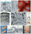
Attributes, differential regulation, and disease association of keratins. (A) Tripartite domain structure shared by all keratin and other intermediate filament (IF) proteins. A central α-helical “rod” domain acts as a key determinant of self-assembly and is flanked by nonhelical “head” and “tail” domains at the N-terminus and C-terminus, respectively. The ends of the rod domain contain 15–20 amino acid regions, here shown is yellow that are highly conserved among all IFs. (B) Visualization of filaments, reconstituted in vitro from purified K5 and K14, by negative staining and electron microscopy. Bar, 125 nm. (C) Ultrastructure of the cytoplasm of epidermal cells in primary culture as shown by transmission electron microscopy. Keratin filaments are abundant and tend to be organized in large bundles of loosely packed filaments in the cytoplasm. Bar, 5 μm. (D) Triple-labeling for keratin (red) and desmoplakin (green), a desmosome component, and DNA (blue) by indirect immunofluorescence of epidermal cells in culture. Keratin filaments are organized in a network that spans the entire cytoplasm and are attached to desmosomes at points of cell–cell contacts (arrowheads). Bar, 30 μm. N, nucleus. (E) Histological cross section of resin-embedded human trunk epidermis, revealing the basal (B), spinous (S), granular (G), and cornified (C) compartments. The differentiation-dependent distribution of keratin proteins in the epidermis is indicated. Bar, 50 μm. N, nucleus. (F) Ultrastructure of the boundary between the basal and suprabasal cells in mouse trunk epidermis as seen by routine transmission electron microscopy. The sample, from which this micrograph was taken, is oriented in the same manner as (E). Organization of keratin filaments as loose bundles (brackets in basal cell) correlates with the expression of K5–K14 in basal cells, whereas the formation of much thicker and electron-dense filament bundles (brackets in spinous cell) reflects the onset of K1–K10 expression in early differentiating keratinocytes. Arrowheads point to desmosomes. Bar, 1 μm. N, nucleus. (G and H) Differential distribution of keratin epitopes on human skin tissue cross sections (similar to E) as visualized by an antibody-based detection method. K14 occurs in the basal layer, where the epidermal progenitor cells reside (G). K10 primarily occurs in the differentiating suprabasal layers of epidermis (H). Dashed line, basal lamina. Bar, 100 μm. (I) Newborn mouse littermates. The top mouse is transgenic (Tg) and expresses a mutated form of K14 in its epidermis. Unlike the control pup below (Wt), this transgenic newborn shows extensive blistering of its front paws (arrows). (J and K) Hematoxylin and eosin (H&E)-stained histological cross section through paraffin-embedded newborn mouse skin similar to those shown in (I). Compared with the intact skin of a control littermate (K, Wt), the epidermis of the K14 mutant expressing transgenic pup (J, Tg) shows intraepidermal cleavage within the basal layer, where the mutant keratin is expressed (opposing arrows). Bar, 100 μm. (L) Leg skin in a patient with the Dowling-Meara form of epidermolysis bullosa simplex. Several skin blisters are grouped in a herpetiform pattern. Reproduced from Coulombe & Bernot, 2004.
Figure 3
Figure 3
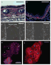
Various analyses of skin keratins utilizing mouse tissue and cultured primary keratinocytes. (A) Hematoxylin and eosin stain of fresh-frozen adult mouse ear tissue (4 months old). Dotted line marks the boundary between the epidermis (Epi) and dermis (Derm). Bar, 50 μm. (B) Fresh-frozen front paw tissue of a 2-month-old mouse processed for immunofluorescence of basal keratin K5. Note the restriction of K5 to the basal (progenitor) layer of keratinocytes. Dotted line marks the boundary between the epidermis (Epi) and dermis (Derm). Bar, 50 μm. (C) Live cell images of “wounding assay” using WT mouse keratinocytes in primary culture. Freshly isolated keratinocytes were plated in chamber slides with culture inserts. The “wound” was introduced by removing culture inserts when cells were 100% confluent. Phase contrast imaging was performed with a Zeiss Axio Observer Z1 microscope equipped with Zeiss EC Plan-Neofluar 10×/0.3 Ph1 objective for 16 h. Bar, 200 μm. (D) Transient expression of mCherry fluorescence protein (mCherry)-tagged paxillin in mouse primary keratinocytes. Freshly isolated keratinocytes were transfected with a plasmid encoding mCherry-tagged paxillin using the nucleofection method before plating in chamber slides with culture inserts. After removing the culture inserts, keratinocytes were allowed to migrate for at least 8 h before imaging using a Zeiss Axio Observer Z1 fluorescence microscope equipped with Zeiss EC Plan-Neofluar 40× objective. Keratinocytes shown in this panel were located at the leading edge in “wounding assay.” Arrow points to a paxillin-positive focal adhesion. Bar, 20 μm. N, nucleus. (E) Immunofluorescence staining of keratin K14 in mouse skin keratinocytes in primary culture. Keratinocytes were isolated from newborn mouse pups and cultured in mKer media for 2 days. They were then fixed with 4% PFA and permeabilized with 0.5% Triton/PBS. Bar, 50 μm. N, nucleus.
Figure 4
Figure 4
Subcellular fractionation methods and assays for studying keratin-interacting proteins. (A) Western blot analysis of Src activity (Y416 phosphorylated epitope) in WT, Krt6a/b+/−, and Krt6a/b−/− skin explant keratinocyte protein lysates. Cellular outgrowths from skin explants cultured for 6 days were pooled and solubilized with RIPA buffer, and then with 6.5 M Urea buffer. Use of an antibody to K14 for western blotting reveals the relative amount of keratin protein occurring at the RIPA and urea extraction steps. β-Actin was used as a loading control for both RIPA-soluble fraction and RIPA-insoluble fraction (urea-soluble fraction). (B) Western blot analysis of subcellular localization of Rac1 in WT, Krt6a/b+/−, and Krt6a/b−/− skin explant keratinocyte protein lysates. Cellular outgrowths from skin explants cultured for 6 days were pooled and solubilized with 0.01% Digitonin buffer and then with 0.5% Triton X-100 buffer. Loading was assessed using β-actin (digitonin-soluble fraction/cytosolic fraction) and caveolin (triton-soluble fraction/membrane fraction). Use of an antibody to K14 for western blotting reveals the relative amount of keratin protein occurring at the RIPA and urea extraction steps. (C) Immunoprecipitation (IP) of keratin 16 (K16) from mouse keratinocytes in primary culture. Keratinocytes were isolated from newborn mouse pups and cultured to confluence before IP. “Input” represents the whole cell lysate, which serves as a positive control, “K16” refers to the K16 antibody (see Table 1) used for IP, and “PIS” refers to preimmune serum, used as a negative control. (D) Far-western assay to study the (direct) interaction between keratin proteins and, in this case, Src protein. Recombinant K5, K6, and K17 proteins were purified using HiTrap Q column. 5 μg of each of these keratin proteins were run on 10% SDS-PAGE and transferred to a nitrocellulose membrane. Ponceau staining was done to assess loading of these proteins. The membrane was then incubated with recombinant Src protein (150 ng/mL; Abcam) for 4 h at room temperature. The association of Src protein with keratin proteins on the nitrocellulose membrane was next detected via conventional western blotting for Src protein. See Rotty and Coulombe (2012), for details.

References

    1. Alvarado DM, Coulombe PA. Directed expression of a chimeric type II keratin partially rescues keratin 5-null mice. The Journal of Biological Chemistry. 2014;289:19435–19447. - PMC - PubMed
    1. Bernot KM, Coulombe PA, McGowan KM. Keratin 16 expression defines a subset of epithelial cells during skin morphogenesis and the hair cycle. The Journal of Investigative Dermatology. 2002;119:1137–1149. - PubMed
    1. Bernot KM, Coulombe PA, Wong P. Skin: An ideal model system to study keratin genes and proteins. Methods in Cell Biology. 2004;78:453–487. - PubMed
    1. Bonifas JM, Rothman AL, Epstein EH., Jr Epidermolysis bullosa simplex: Evidence in two families for keratin gene abnormalities. Science. 1991;254:1202–1205. - PubMed
    1. Byrne C. Regulation of gene expression in developing epidermal epithelia. Bioessays. 1997;19:691–698. - PubMed

Publication types

LinkOut - more resources