

Survival of irradiated recipient mice after transplantation of bone marrow from young, old and "early aging" mice
- PMID: 26796640
- PMCID: PMC4712343
- DOI: 10.18632/aging.100867
Survival of irradiated recipient mice after transplantation of bone marrow from young, old and "early aging" mice
Abstract
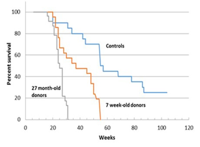
Bone marrow transplantation is used to examine survival, hematopoietic stem cell function and pathology in recipients of young and old wild type bone marrow derived stem cells (BMDSCs) as well as cells from p53-based models of premature aging. There is no difference in the long term survival of recipients of 8 week-old p53+/m donor cells compared to recipients of 8 week-old wild-type (WT) donor cells (70 weeks) or of recipients of 16-18 weeks-old donor cells from either p53+/m or WT mice. There is shorter survival in recipients of older versus younger WT donor bone marrow, but the difference is only significant when comparing 8 and 18 week-old donors. In the p44-based model, short term survival/engraftment is significantly reduced in recipients of 11 month-old p44 donor cells compared to 4 week-old p44 or wild type donor cells of either age; mid-life survival at 40 weeks is also significantly less in recipients of p44 cells. BMDSCs are readily detectable within recipient bone marrow, lymph node, intestinal villi and liver sinusoids, but not in epithelial derived cells. These results indicate that recipients of young BMDSCs may survive longer than recipients of old bone marrow, but the difference is marginal at best.
Keywords: aging; bone marrow transplantation; p44; p53.
Conflict of interest statement
The authors declare that they have no conflicts of interest.
Figures


Similar articles
-
[Influence of donor mouse age on the establishment of murine acute graft versus host disease model after allogenic hematopoietic stem cell transplantation].Zhongguo Shi Yan Xue Ye Xue Za Zhi. 2014 Oct;22(5):1365-70. doi: 10.7534/j.issn.1009-2137.2014.05.035. Zhongguo Shi Yan Xue Ye Xue Za Zhi. 2014. PMID: 25338590 Chinese.
-
The contribution of cytotoxic and noncytotoxic function by donor T-cells that support engraftment after allogeneic bone marrow transplantation.Biol Blood Marrow Transplant. 2002;8(11):588-96. doi: 10.1053/bbmt.2002.v8.abbmt080588. Biol Blood Marrow Transplant. 2002. PMID: 12463477
-
Expansion of hematopoietic stem cell phenotype and activity in Trp53-null mice.Exp Hematol. 2003 Jun;31(6):521-7. doi: 10.1016/s0301-472x(03)00072-9. Exp Hematol. 2003. PMID: 12829028
-
Unrelated donor bone marrow transplantation for hematological malignancies-current status.Leuk Lymphoma. 1996 Oct;23(3-4):221-6. doi: 10.3109/10428199609054824. Leuk Lymphoma. 1996. PMID: 9031102 Review.
-
Bone marrow transplantation: a new strategy for intractable diseases.Drugs Today (Barc). 2002 Feb;38(2):103-11. doi: 10.1358/dot.2002.38.2.820106. Drugs Today (Barc). 2002. PMID: 12532188 Review.
Cited by
-
Animal Welfare in Radiation Research: The Importance of Animal Monitoring System.Vet Sci. 2023 Nov 10;10(11):651. doi: 10.3390/vetsci10110651. Vet Sci. 2023. PMID: 37999474 Free PMC article. Review.
-
Inducible disruption of the c-myb gene allows allogeneic bone marrow transplantation without irradiation.J Immunol Methods. 2018 Jun;457:66-72. doi: 10.1016/j.jim.2018.03.016. Epub 2018 Apr 6. J Immunol Methods. 2018. PMID: 29630967 Free PMC article.
-
Mobilization-based transplantation of young-donor hematopoietic stem cells extends lifespan in mice.Aging Cell. 2020 Mar;19(3):e13110. doi: 10.1111/acel.13110. Epub 2020 Feb 3. Aging Cell. 2020. PMID: 32012439 Free PMC article.
-
Reducing functionally defective old HSCs alleviates aging-related phenotypes in old recipient mice.Cell Res. 2025 Jan;35(1):45-58. doi: 10.1038/s41422-024-01057-5. Epub 2025 Jan 2. Cell Res. 2025. PMID: 39743633 Free PMC article.
References
-
- Jacobson LO, Simmons EL, Marks EK, Robson MJ, Bethard WF, Gaston EO. The role of the spleen in radiation injury and recovery. J Lab Clin Med. 1950;35:746–770. - PubMed
-
- Lorenz E, Uphoff D, Reid TR, Shelton E. Modification of irradiation injury in mice and guinea pigs by bone marrow injections. J Natl Cancer Inst. 1951;12:197–201. - PubMed
-
- Harrison DE. Mouse erythropoietic stem cell lines function normally 100 months: loss related to number of transplantations. Mech Ageing Dev. 1979;9:427–433. - PubMed
Publication types
MeSH terms
Substances
Grants and funding
LinkOut - more resources
Full Text Sources
Other Literature Sources
Medical
Research Materials
Miscellaneous
