. 2016 Jan 22:6:19839.
doi: 10.1038/srep19839.

Membrane omega-3 fatty acids modulate the oligomerisation kinetics of adenosine A2A and dopamine D2 receptors

Affiliations

Membrane omega-3 fatty acids modulate the oligomerisation kinetics of adenosine A2A and dopamine D2 receptors

Ramon Guixà-González et al. Sci Rep. .

Abstract

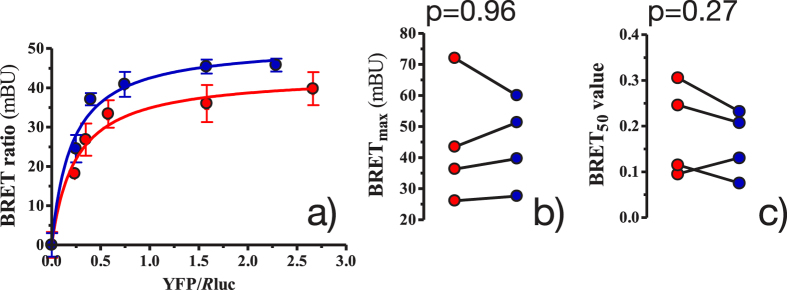
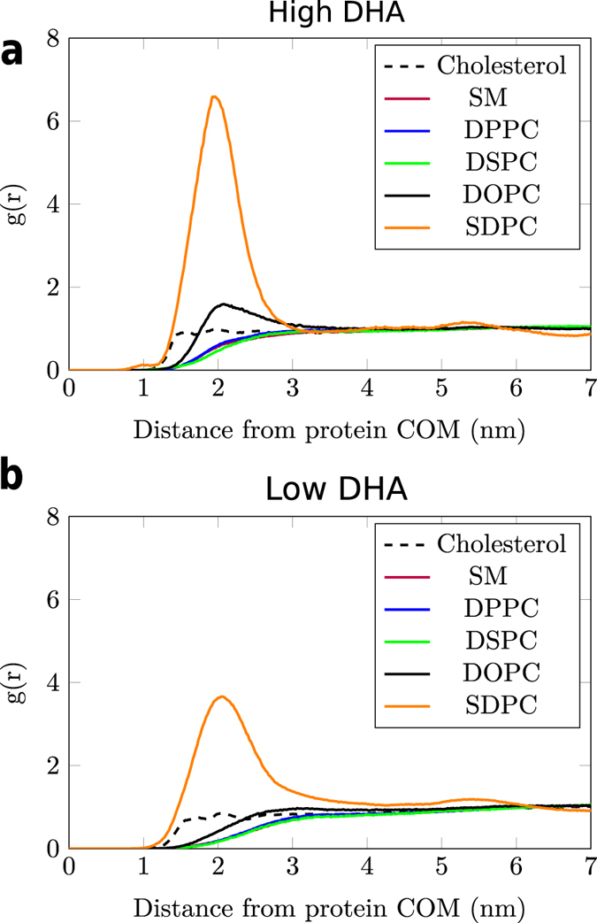
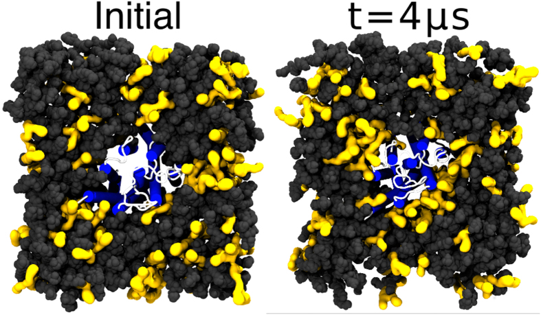
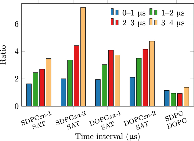
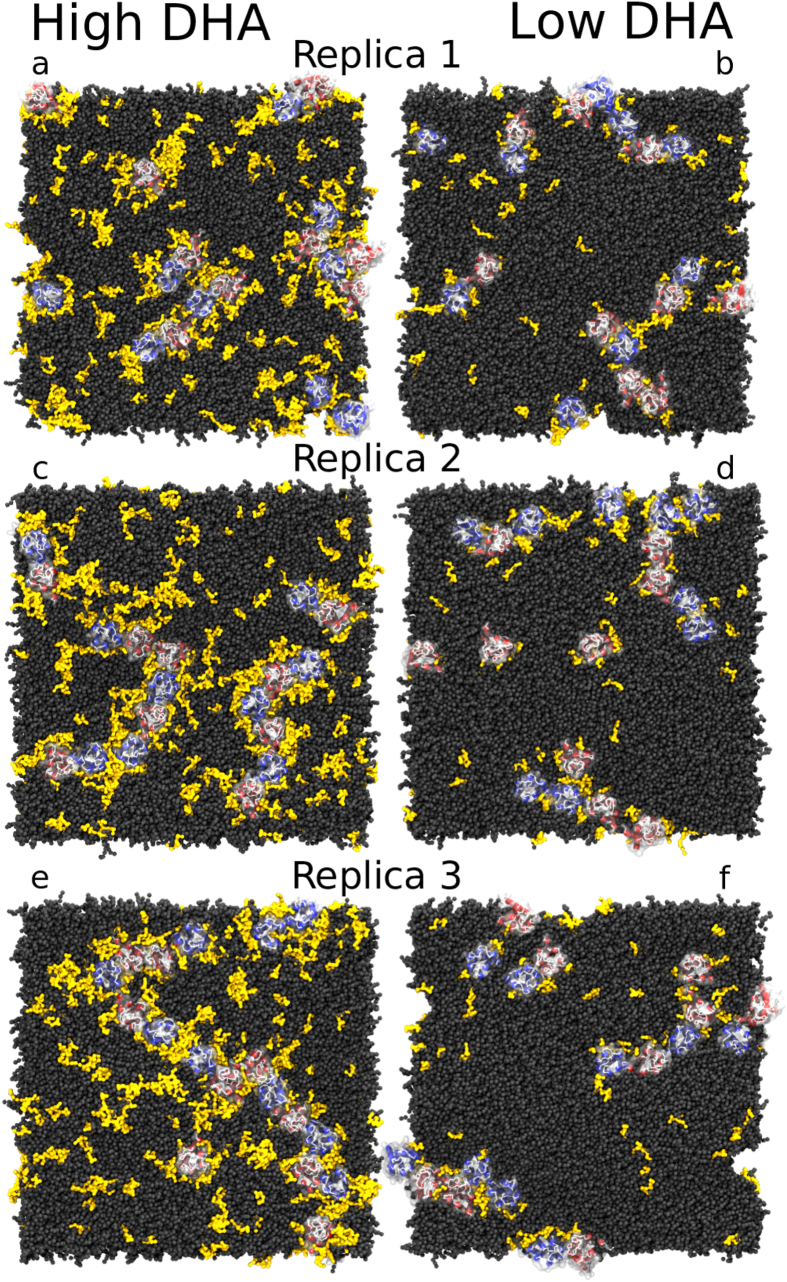
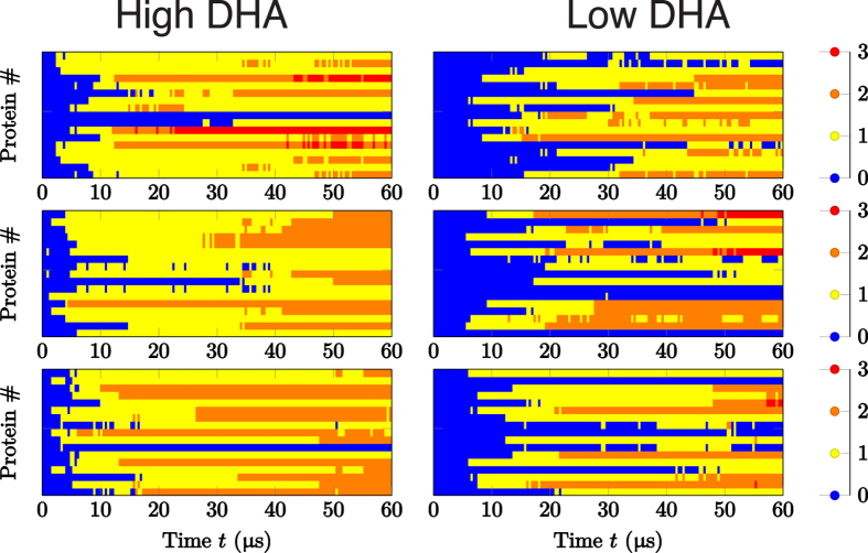
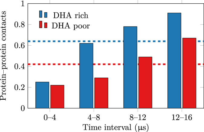
Membrane levels of docosahexaenoic acid (DHA), an essential omega-3 polyunsaturated fatty acid (ω-3 PUFA), are decreased in common neuropsychiatric disorders. DHA modulates key cell membrane properties like fluidity, thereby affecting the behaviour of transmembrane proteins like G protein-coupled receptors (GPCRs). These receptors, which have special relevance for major neuropsychiatric disorders have recently been shown to form dimers or higher order oligomers, and evidence suggests that DHA levels affect GPCR function by modulating oligomerisation. In this study, we assessed the effect of membrane DHA content on the formation of a class of protein complexes with particular relevance for brain disease: adenosine A2A and dopamine D2 receptor oligomers. Using extensive multiscale computer modelling, we find a marked propensity of DHA for interaction with both A2A and D2 receptors, which leads to an increased rate of receptor oligomerisation. Bioluminescence resonance energy transfer (BRET) experiments performed on living cells suggest that this DHA effect on the oligomerisation of A2A and D2 receptors is purely kinetic. This work reveals for the first time that membrane ω-3 PUFAs play a key role in GPCR oligomerisation kinetics, which may have important implications for neuropsychiatric conditions like schizophrenia or Parkinson's disease.


Figures

Figure 1
Figure 1. Effect of DHA on the amount of A2A–D2 heteromerisation in cellular steady state with low (red) and high (blue) DHA content.
(a) shows representative BRET saturation curves, where each point measurement was performed in triplicate. BRET ratios (×1000) are in mBRET units (mBU). Error bars show the SEM. (b,c) columns show, respectively, the BRETmax and BRET50 results of 4 independent experiments with low (red) and high (blue) amounts of DHA. These results were compared by a paired t test and the p values are shown.
Figure 2
Figure 2
Probability density (i.e. radial distribution function, g(r)) of lipids around the center of mass (COM) of the A2A receptor embedded in healthy- (high DHA, (a)) and diseased-like (low DHA, (b)) model membranes in CG-MD simulations. y axis represent g(r) (arbitrary units) and x axis the distance to the protein COM in nm. The radial distribution function of SM heavily overlaps with the rest of saturated lipids (i.e. DPPC and DSPC). Radial distribution functions for the D2 receptor are shown in Fig. S4.
Figure 3
Figure 3. Initial and final (4 μs) snapshots of the all-atom simulation of the A2A receptor embedded in a healthy-like membrane (high DHA).
Protein helices are depicted in blue and loops in white. Unsaturated phospholipids (SDPC and POPC) are drawn as yellow surfaces with dark grey spheres corresponding to other lipid types. Water and ions are omitted for clarity.
Figure 4
Figure 4. Ratios of A2A receptor–lipid contacts of unsaturated (SDPC and POPC) and saturated (SAT) lipid tails during the all-atom simulation.
Figure 5
Figure 5. Final snapshots of healthy- and diseased-like systems after 60 μs of CG-MD simulation.
Left and right columns display 3 replicas of healthy- (high DHA, left) and diseased-like (low DHA, right) systems. A2A and D2 helices are depicted in red and blue cartoons, respectively. Dark grey spheres correspond to all membrane lipids except for SDPC molecules depicted in yellow surface. Water and ions are not shown for clarity.
Figure 6
Figure 6. Time-dependence of protein aggregation in CG-MD simulations.
Data are shown for healthy-like (i.e. high DHA, left) and diseased-like (low DHA, right) systems. Each cell represents one of the three replicas and each line in the plots corresponds to an individual receptor. The colour code reflects the number of contacts per protomer. Corresponding data for the short (16 μs) systems is shown in Fig. S9.
Figure 7
Figure 7. Time evolution of the mean number of protein–protein contacts per protomer in the set of shorter simulations.
‘Healthy’ and ‘Diseased’ refer to healthy-like (high DHA) and diseased-like (low DHA) model membranes. Average values are shown by dashed lines.

References

    1. Taha A. Y., Cheon Y., Ma K., Rapoport S. I. & Rao J. S. Altered fatty acid concentrations in prefrontal cortex of schizophrenic patients. J. Psychiat. Res. 47, 636–643 (2013). - PMC - PubMed
    1. Martín V. et al. Lipid alterations in lipid rafts from Alzheimer’s disease human brain cortex. J. Alzheimers Dis. 19, 489–502 (2010). - PubMed
    1. Fabelo N. et al. Severe alterations in lipid composition of frontal cortex lipid rafts from Parkinson’s disease and incidental Parkinson’s disease. Mol. Med. 17, 1107–1118 (2011). - PMC - PubMed
    1. Innis S. M. Dietary omega 3 fatty acids and the developing brain. Brain Res. 1237, 35–43 (2008). - PubMed
    1. Calon F. & Cole G. Neuroprotective action of omega-3 polyunsaturated fatty acids against neurodegenerative diseases: evidence from animal studies. Prostag. Leukotr. Ess. 77, 287–293 (2007). - PubMed

Publication types