. 2016 Jan 21;11(1):e0147447.
doi: 10.1371/journal.pone.0147447. eCollection 2016.

Modeling Staphylococcus epidermidis-Induced Non-Unions: Subclinical and Clinical Evidence in Rats

Affiliations

Modeling Staphylococcus epidermidis-Induced Non-Unions: Subclinical and Clinical Evidence in Rats

Arianna Barbara Lovati et al. PLoS One. .

Abstract

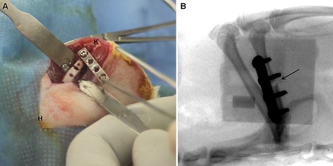
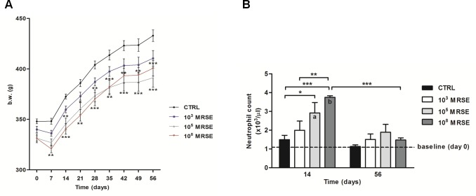
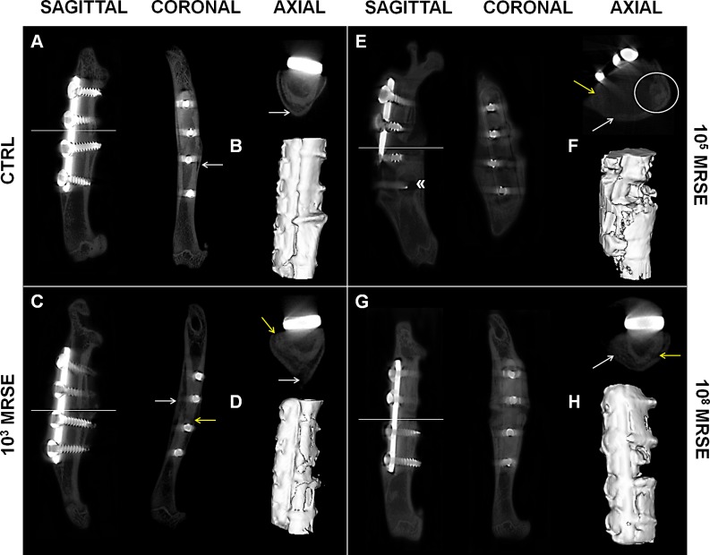
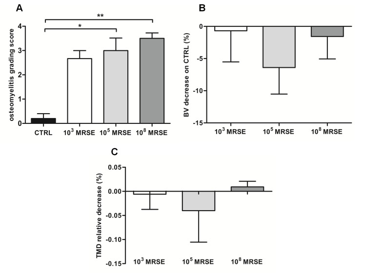
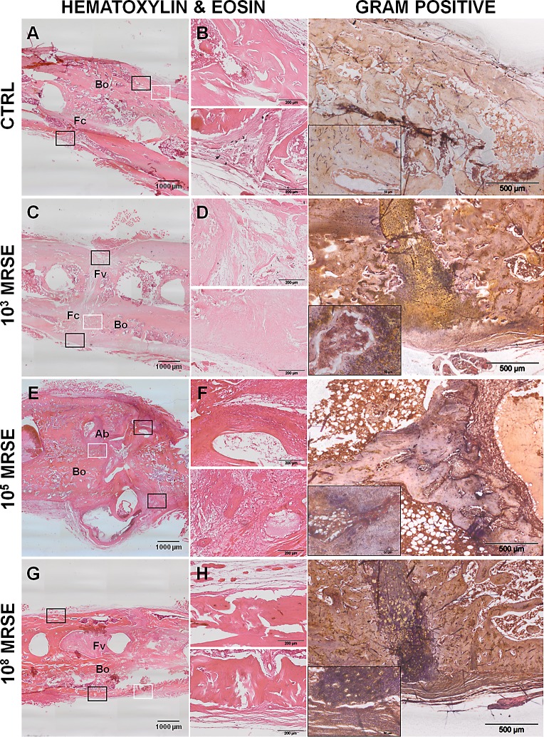
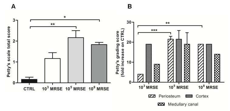
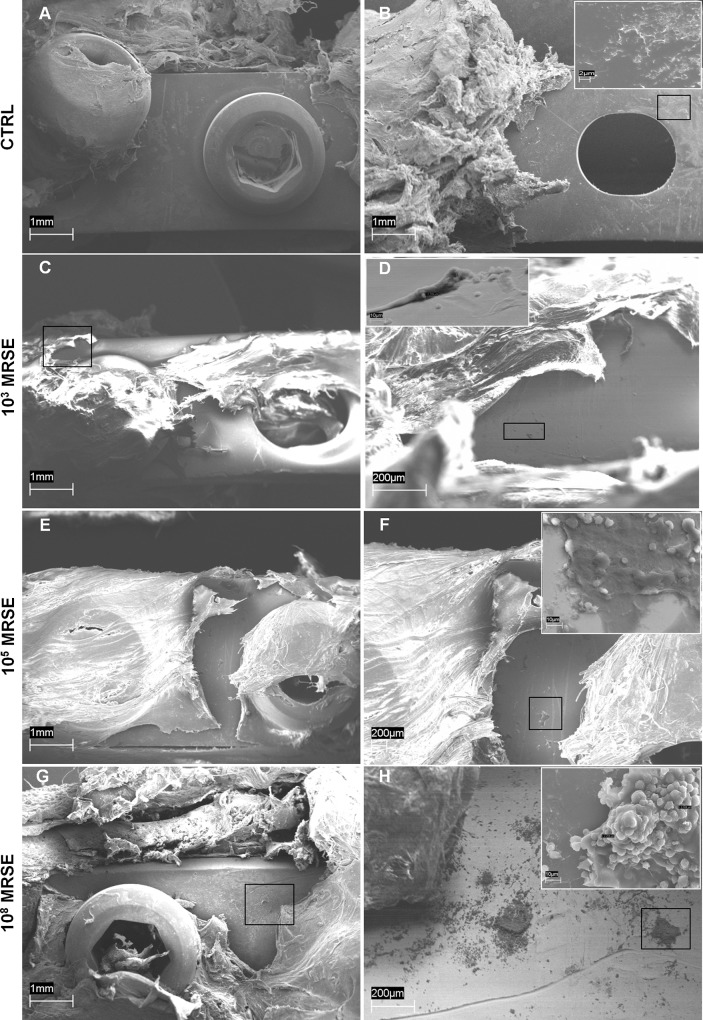
S. epidermidis is one of the leading causes of orthopaedic infections associated with biofilm formation on implant devices. Open fractures are at risk of S. epidermidis transcutaneous contamination leading to higher non-union development compared to closed fractures. Although the role of infection in delaying fracture healing is well recognized, no in vivo models investigated the impact of subclinical low-grade infections on bone repair and non-union. We hypothesized that the non-union rate is directly related to the load of this commonly retrieved pathogen and that a low-grade contamination delays the fracture healing without clinically detectable infection. Rat femurs were osteotomized and stabilized with plates. Fractures were infected with a characterized clinical-derived methicillin-resistant S. epidermidis (10(3), 10(5), 10(8) colony forming units) and compared to uninfected controls. After 56 days, bone healing and osteomyelitis were clinically assessed and further evaluated by micro-CT, microbiological and histological analyses. The biofilm formation was visualized by scanning electron microscopy. The control group showed no signs of infection and a complete bone healing. The 10(3) group displayed variable response to infection with a 67% of altered bone healing and positive bacterial cultures, despite no clinical signs of infection present. The 10(5) and 10(8) groups showed severe signs of osteomyelitis and a non-union rate of 83-100%, respectively. The cortical bone reaction related to the periosteal elevation in the control group and the metal scattering detected by micro-CT represented limitations of this study. Our model showed that an intra-operative low-grade S. epidermidis contamination might prevent the bone healing, even in the absence of infectious signs. Our findings also pointed out a dose-dependent effect between the S. epidermidis inoculum and non-union rate. This pilot study identifies a relevant preclinical model to assess the role of subclinical infections in orthopaedic and trauma surgery and to test specifically designed diagnostic, prevention and therapeutic strategies.


Conflict of interest statement

Competing Interests: The authors have declared that no competing interests exist.

Figures

Fig 1
Fig 1. Plate positioning and postoperative analysis.
(A) Plate diversion from the femur and the creation of a 1 mm non-critical midshaft full-thickness defect. The anatomical sites are reported as knee (K) and hip (H) joints. (B) Fluoroscopic examination of the femoral fracture and the correct plate position. The fracture of the femoral midshaft is shown by a black arrow.
Fig 2
Fig 2. Clinical data.
(A) The graph shows the numerical values of body weight (g) in the experimental groups over time. (B) The histogram shows the neutrophil count among experimental groups at day 14 and 56 after surgery. The dotted line represents the baseline at day 0 before surgery. Comparisons between groups and time points were analyzed with two-way ANOVA and Bonferroni’s post-hoc. Statistical significance was P<0.05 (*, a, b), P<0.01 (**) and P<0.001 (***), n = 6.
Fig 3
Fig 3. Qualitative micro-CT imaging and isosurface.
The representative panel shows micro-CT images on the day of explantation. The sagittal, coronal and axial planes (A, C, E, G), as well as a three-dimensional isosurface reconstruction (B, D, F, H) were presented for the control (CTRL), and the 103, 105, and 108 MRSE groups. Symbols indicate: cortical reaction (white arrows); loss of cortical wall and osteolysis (yellow arrows); peri-implant osteolysis («); presence of abscesses (white circle).
Fig 4
Fig 4. Micro-CT-based semiquantitative and quantitative analyses of bone structure.
(A) Osteomyelitis grading score based on Odekerken’s scale. (B) Bone volume (BV) quantitative analysis of the infected groups normalized on the control group, reported as a percentage. (C) Tissue mineral density (TMD) quantitative analysis of the infected groups normalized on the control group, reported as a percentage. Comparisons among groups were analyzed by one-way ANOVA. Statistical significance for P<0.05 (*), n = 6.
Fig 5
Fig 5. Histological analysis at the day of explantation.
The representative panel shows H&E and Gram staining in the four groups. The panels depict an overview of the samples, magnification 2x, scale bar 1000μm (A, C, E, G). The panels depict areas of interest of the black boxes, magnification 10x, scale bar 200μm (B, D, F, H). The Gram staining depicts bacterial colonies identified by the white boxes, magnification 4x, scale bar 500μm and specific regions within the small boxes, magnification 40x, scale bar 50μm. Bo, bone; Fc, fibrocartilaginous callus; Fv, fibrovascular tissue; Ab, abscesses.
Fig 6
Fig 6. Histological score based on Petty’s scale.
The histogram compares the semiquantitative score performed on the periosteum, cortex and medullary canal regarding the osteomyelitis signs. (A) Total score analysis. (B) Fold increase of the grading scale of the infected groups with respect to the control group. Comparisons among groups were analyzed by one-way ANOVA. Statistical significance for P<0.05 (*), P<0.01 (**), P<0.001 (***), n = 5.
Fig 7
Fig 7. Representative photographs of SEM analysis of biofilm formation.
Absence of biofilm formation in the control group either on the top (A) or on the bottom (B) of the implanted plate (magnification 40x, scale bar 1mm) with the presence of bony bridging structure on the bone-implant interface (B small box, magnification 6700x, scale bar 2μm). Presence of few coccidal bacteria on the top of the plate (C, D, magnifications 40x and 250x, scale bars 1mm and 200μm, respectively) occasionally coated with mucoid material in the 103MRSE group (D small box, magnification 3000x scale bar 10μm). Presence of cocci within the peri-implant fibrous tissues covering the plate and on the top of the plate (E, magnification 40x, scale bar 1mm), free or partially embedded into mucous-gelatinous matrix in the 105MRSE group (F, magnification 70x, scale bar 200μm; F small box, magnification 3000x scale bar 10μm). Clusters of cocci adhering to the top of the plate surface (G, H, magnifications 40x and 90x, scale bars 1mm and 200μm, respectively) completely embedded in a well-organized biofilm matrix in the 108MRSE group (H small box, magnification 3000x, scale bar 10μm).

References

    1. Alt V, Lips KS, Henkenbehrens C, Muhrer D, Oliveira Cavalcanti MC, Sommer U, et al. A new animal model for implant-related infected non-unions after intramedullary fixation of the tibia in rats with fluorescent in situ hybridization of bacteria in bone infection. Bone 2011;48:1146–1153. 10.1016/j.bone.2011.01.018 - DOI - PubMed
    1. Trampuz A, Zimmerli W. Diagnosis and treatment of implant associated septic arthritis and osteomyelitis. Curr Infect Dis Rep 2008;10:394–403. - PubMed
    1. Kim PH, Leopold SS. Gustilo-Anderson classification. Clin Orthop Relat Res 2012;470:3270–3274. 10.1007/s11999-012-2376-6 - DOI - PMC - PubMed
    1. Ostermann PA, Henry SL, Seligson D. Timing of wound closure in severe compound fractures. Orthopedics 1994;17:397–399. - PubMed
    1. Seligson D, Klemm K. Adult posttraumatic osteomyelitis of the tibial diaphysis of the tibial shaft. Clin Orthop Relat Res 1999;360:30–36. - PubMed

Publication types

MeSH terms