. 2016 Mar 18;291(12):6218-31.
doi: 10.1074/jbc.M115.710707. Epub 2016 Jan 21.

The Ankrd13 Family of Ubiquitin-interacting Motif-bearing Proteins Regulates Valosin-containing Protein/p97 Protein-mediated Lysosomal Trafficking of Caveolin 1

Affiliations

The Ankrd13 Family of Ubiquitin-interacting Motif-bearing Proteins Regulates Valosin-containing Protein/p97 Protein-mediated Lysosomal Trafficking of Caveolin 1

Daocharad Burana et al. J Biol Chem. .

Abstract

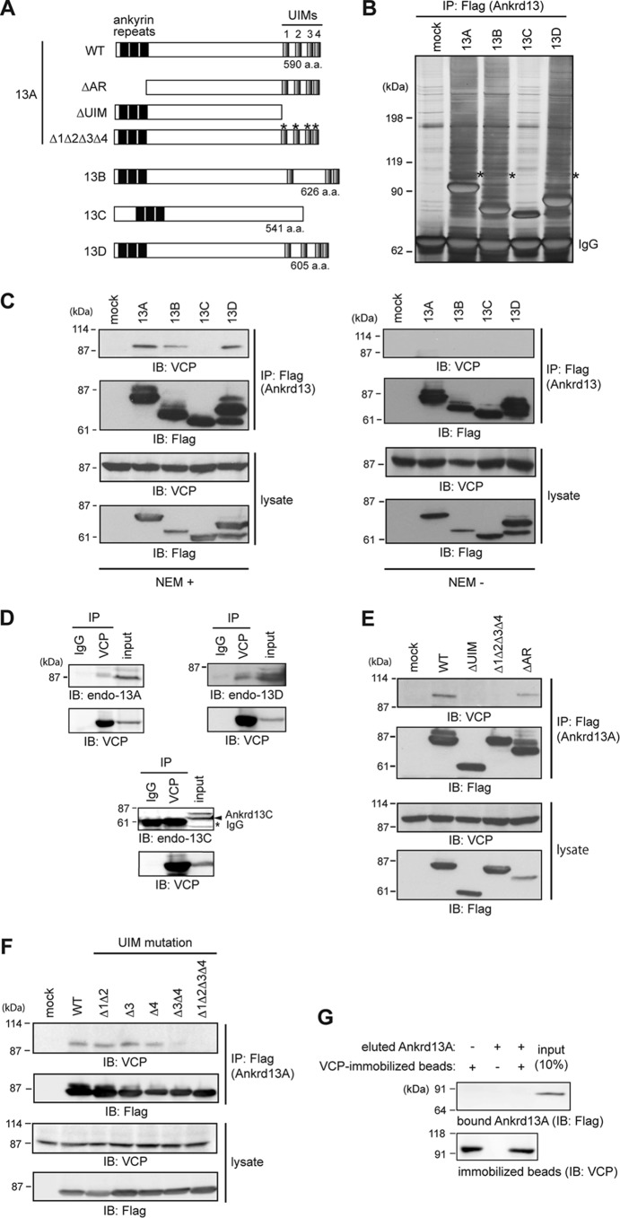
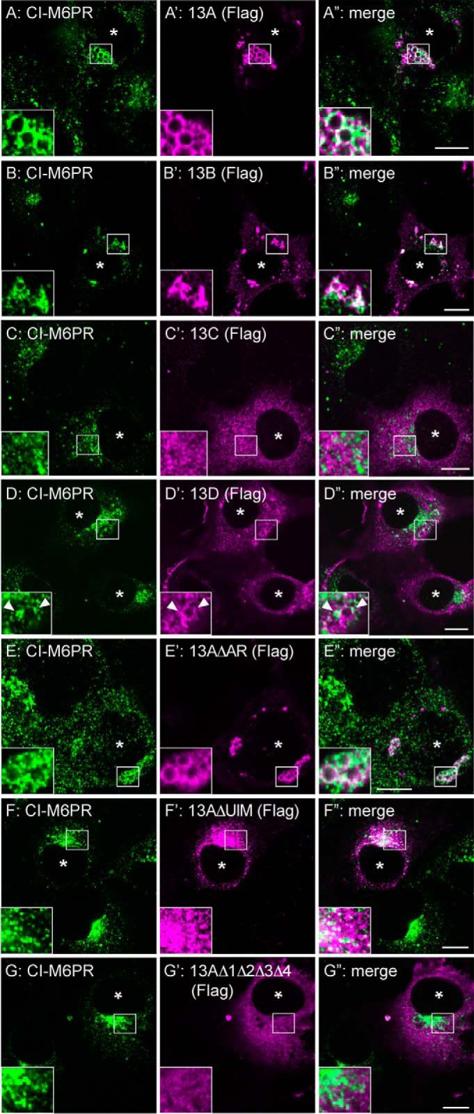
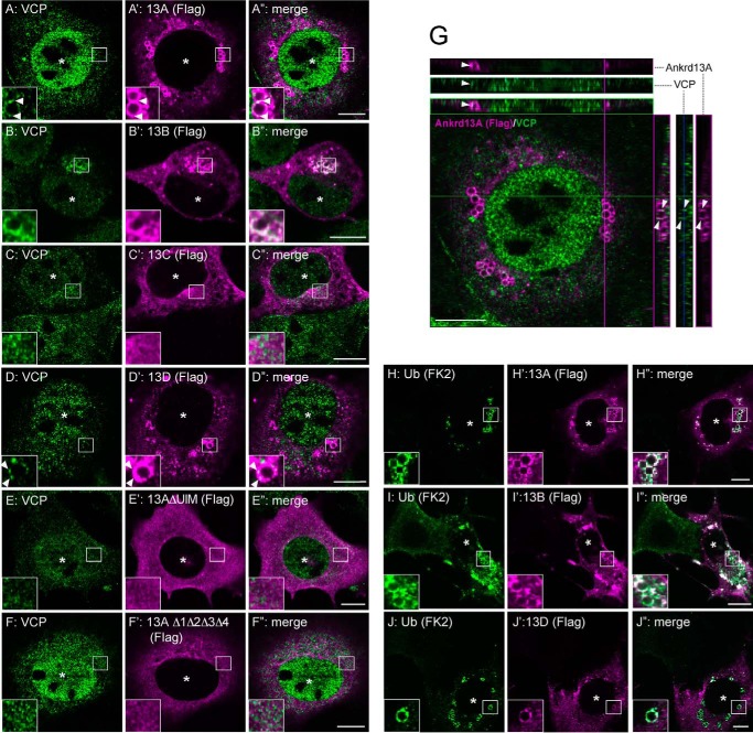
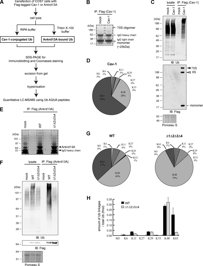
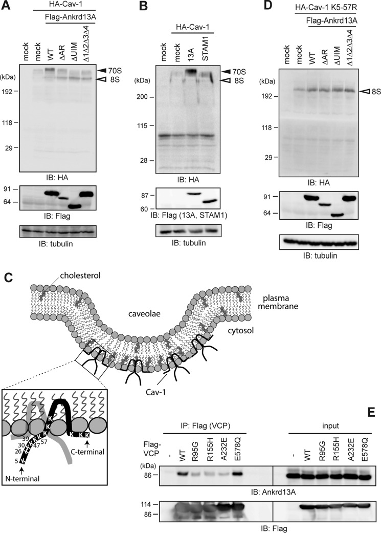
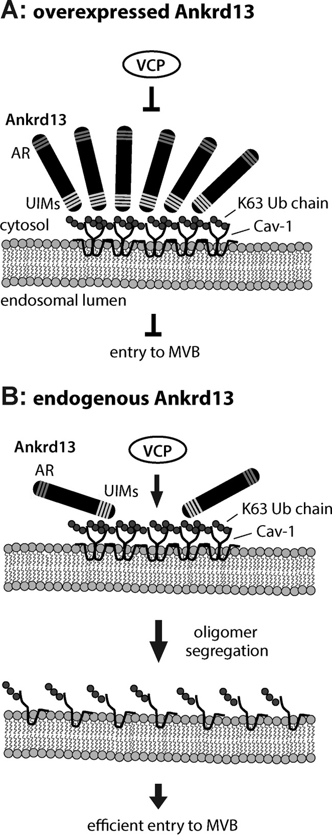
Caveolin 1 (Cav-1) is an oligomeric protein that forms flask-shaped, lipid-rich pits, termed caveolae, on the plasma membrane. Cav-1 is targeted for lysosomal degradation in ubiquitination- and valosin-containing protein (VCP)-dependent manners. VCP, an ATPase associated with diverse cellular activities that remodels or segregates ubiquitinated protein complexes, has been proposed to disassemble Cav-1 oligomers on the endosomal membrane, facilitating the trafficking of Cav-1 to the lysosome. Genetic mutations in VCP compromise the lysosomal trafficking of Cav-1, leading to a disease called inclusion body myopathy with Paget disease of bone and/or frontotemporal dementia (IBMPFD). Here we identified the Ankrd13 family of ubiquitin-interacting motif (UIM)-containing proteins as novel VCP-interacting molecules on the endosome. Ankrd13 proteins formed a ternary complex with VCP and Cav-1 and exhibited high binding affinity for ubiquitinated Cav-1 oligomers in an UIM-dependent manner. Mass spectrometric analyses revealed that Cav-1 undergoes Lys-63-linked polyubiquitination, which serves as a lysosomal trafficking signal, and that the UIMs of Ankrd13 proteins bind preferentially to this ubiquitin chain type. The overexpression of Ankrd13 caused enlarged hollow late endosomes, which was reminiscent of the phenotype of the VCP mutations in IBMPFD. Overexpression of Ankrd13 proteins also stabilized ubiquitinated Cav-1 oligomers on the limiting membrane of enlarged endosomes. The interaction with Ankrd13 was abrogated in IMBPFD-associated VCP mutants. Collectively, our results suggest that Ankrd13 proteins cooperate with VCP to regulate the lysosomal trafficking of ubiquitinated Cav-1.

Keywords: VCP/p97; caveolin; cell surface protein; endocytosis; endosome; membrane trafficking; protein degradation; ubiquitin; ubiquitin-interacting motif; ubiquitylation (ubiquitination).


Figures

FIGURE 1.
FIGURE 1.
Ankrd13 proteins interact with VCP. A, schematics of the Ankrd13A-13D and Ankrd13A mutants used routinely in this study. a.a., amino acids. B, HeLa cells were transfected with FLAG-tagged Ankrd13 proteins and lysed in the presence of NEM. Ankrd13 proteins were immunoprecipitated (IP) with an anti-FLAG antibody, and the precipitated proteins were detected by silver staining after SDS-PAGE. Asterisks, the band corresponding to VCP; IgG, IgG heavy chain used for immunoprecipitation. C, HeLa cells transfected with FLAG-tagged Ankrd13 proteins were lysed in the presence (left panel) or absence (right panel) of NEM. Lysates were immunoprecipitated with an anti-FLAG antibody and immunoblotted (IB) with the indicated antibodies. Cell lysates were also immunoblotted with the indicated antibodies. D, the lysate of untransfected HeLa cells, prepared in the presence of NEM, was immunoprecipitated with an anti-VCP antibody and immunoblotted with antibodies to Ankrd13A, 13C, or 13D. E and F, HeLa cells transfected with the indicated FLAG-Ankrd13A mutants were lysed in the presence of NEM, immunoprecipitated with an anti-FLAG antibody, and immunoblotted with the indicated antibodies. G, COS-7 cells were transfected with FLAG-Ankrd13A and lysed using the hot lysis method. FLAG-Ankrd13A was immunoprecipitated with an anti-FLAG antibody and eluted with the FLAG peptide. VCP was immunopurified similarly from the hot lysis lysate of untransfected COS-7 cells and immobilized on anti-VCP antibody-coupled beads. Eluted FLAG-Ankrd13A was incubated with VCP-immobilized beads, and bound Ankrd13A was examined by anti-FLAG immunoblotting. All experiments were performed at least three times.
FIGURE 2.
FIGURE 2.
Ankrd13 overexpression causes enlarged late endosomes. COS-7 cells were transfected with FLAG-tagged Ankrd13 proteins or Ankrd13A mutants and double-stained with anti-CI-M6PR (A–G) and anti-FLAG (A'–G') antibodies. The arrowheads in D–D” indicate the co-localization sites of Ankrd13D with CI-M6PR. Asterisks indicate nuclei in FLAG-Ankrd13-expressing cells. Insets show higher magnification images of regions indicated by squares. Scale bars = 10 μm. All panels are representatives of four independent experiments.
FIGURE 3.
FIGURE 3.
Ankrd13 overexpression recruits VCP and Ub-protein conjugates to enlarged late endosomes. A–F”, COS-7 cells were transfected with FLAG-Ankrd13 proteins or Ankrd13A mutants and double-stained with anti-VCP (A–F) and anti-FLAG (A'–F') antibodies. G, a representative of the orthogonal reconstruction of 10 confocal slices of Ankrd13A-expressing cells is shown in the y-z and x-z axes. H–J”, COS-7 cells were transfected with FLAG-tagged Ankrd13 proteins and double-stained with anti-Ub (FK2, H–J) and anti-FLAG (H'–J') antibodies. Asterisks indicate nuclei in FLAG-Ankrd13-expressing cells. Arrowheads in A–A”, D–D”, and G indicate the co-localization sites of Ankrd13 proteins with VCP. Insets show higher magnification images of the regions indicated by squares. Scale bar = 10 μm. All panels are representatives of three independent experiments.
FIGURE 4.
FIGURE 4.
Ankrd13 proteins bind to Cav-1. A, lysates of COS-7 cells co-transfected with FLAG-Ankrd13A and HA-Cav-1 were immunoprecipitated (IP) with an anti-FLAG antibody and immunoblotted (IB) with the indicated antibodies. The black arrowhead, white arrowheads, and arrows indicate the unmodified monomeric, ubiquitinated, and oligomeric forms of Cav-1, respectively. B, quantification of the immunoblot data in A, demonstrating the proportion of unmodified monomeric, ubiquitinated, and oligomeric forms of Cav-1 in the Ankrd13A immunoprecipitate (IP: FLAG) and cell lysate. Data are normalized against mock-transfected cells and shown as the mean ± S.D. of three independent experiments. C and D, HA-Cav-1 was transfected into COS-7 cells together with FLAG-tagged Ankrd13 proteins (C) or Ankrd13A mutants (D) and examined by co-immunoprecipitation experiments as in A. The 8S and 70S Cav-1 oligomers were not separated by SDS-PAGE with a high concentration (12%) of acrylamide in A, C, and D. E, the lysate of COS-7 cells transfected with FLAG-VCP, HA-Cav-1, and untagged Ankrd13A was subjected to sequential immunoprecipitation. VCP was first immunoprecipitated with an anti-FLAG antibody (IP1) and then eluted from the antibody with the FLAG peptide. The eluate was then immunoprecipitated with an anti-HA antibody (IP2), and the IP1 and IP2 precipitates were immunoblotted with the indicated antibodies. The experiments in C, D, and E were repeated at least three times.
FIGURE 5.
FIGURE 5.
Identification of Ub chain types conjugated to Cav-1 and bound to Ankrd13A. A, the procedures of sample preparation for the LC-MS/MS of Cav1-conjugated and Ankrd13A-bound Ub chains. RIPA, radio-immunoprecipitation assay; AQUA, absolute quantification. B, the lysates of COS-7 cells transfected with FLAG-Cav-1 were subjected to anti-FLAG immunoprecipitation (IP) followed by SDS-PAGE and Coomassie staining. The regions enclosed by dashed lines were excised and analyzed by LC-MS/MS. C, aliquots of the immunoprecipitates in B were immunoblotted (IB) with the indicated antibodies. D, the percentages of Ub chain types in immunoprecipitated FLAG-Cav-1 in B analyzed by LC-MS/MS. E and F, the lysates of COS-7 cells transfected with FLAG-tagged WT Ankrd13A or the Δ1Δ2Δ3Δ4 mutant were subjected to anti-FLAG immunoprecipitation followed by Coomassie staining (E) and immunoblotting (F). The regions enclosed by dashed lines in E were excised and analyzed by LC-MS/MS. G, the percentages of Ub chain types bound to WT Ankrd13A (left panel) and Δ1Δ2Δ3Δ4 (right panel). All data represent the average values obtained from three independent experiments. H, the absolute amounts of Ub chain types bound to Ankrd13A WT and Δ1Δ2Δ3Δ4 mutant prepared in E and F. Error bars indicate mean ± S.D.
FIGURE 6.
FIGURE 6.
Ankrd13 overexpression recruits Cav-1 to enlarged late endosomes. COS-7 cells were transfected with HA-Cav-1 together with FLAG-tagged Ankrd13 proteins or Ankrd13A mutants and double-stained with anti-HA (A–H) and anti-FLAG (A'–H') antibodies. Insets show higher magnification images of the regions indicated by squares. Scale bars = 10 μm. All panels are representatives of three independent experiments.
FIGURE 7.
FIGURE 7.
Ankrd13A overexpression stabilizes the Cav-1 70S oligomer. A and B, COS-7 cells were transfected with HA-Cav-1 together with FLAG-tagged Ankrd13A or its mutants (A) or FLAG-STAM1 (B). Cell lysates were prepared in low-SDS sample buffer without boiling. The same amounts of total proteins were immunoblotted with the indicated antibodies. IB, immunoblotting. C, schematics of the caveolae and human Cav-1, showing the positions of all Lys residues in Cav-1. Six Lys residues in the N-terminal region (Lys-5 to Lys-57) were replaced by Arg in the K5–57R mutant. D, COS-7 cells were transfected with HA-Cav-1 K5–57R together with FLAG-tagged Ankrd13A or its mutants. Cells were examined as in A. E, COS-7 cells were transfected with FLAG-tagged WT VCP or its mutants identified in patients with IBMPFD (R95G, R155H, and A232E) or lacking ATPase activity (E578Q). Their lysates were immunoprecipitated (IP) with an anti-FLAG antibody, and the precipitates were immunoblotted with the indicated antibodies. All the experiments were performed three times.
FIGURE 8.
FIGURE 8.
Schematics for the Ankrd13 function on endosomal Cav-1 trafficking. A, when overexpressed, Ankrd13 blocks VCP interaction with the Cav-1 oligomer on the endosomal membrane, inhibiting Cav-1 entry into inner vesicles of the MVB. AR, ankyrin repeat. B, endogenous Ankrd13 concentrates ubiquitinated Cav-1 on the endosomal membrane, facilitating VCP-mediated segregation of the Cav-1 oligomer and subsequent entry of Cav-1 into the MVB inner vesicles.

Similar articles

Cited by

References

    1. Parton R. G., and Simons K. (2007) The multiple faces of caveolae. Nat. Rev. Mol. Cell Biol. 8, 185–194 - PubMed
    1. Hayer A., Stoeber M., Bissig C., and Helenius A. (2010) Biogenesis of caveolae: stepwise assembly of large caveolin and cavin complexes. Traffic 11, 361–382 - PubMed
    1. Parton R. G., and del Pozo M. A. (2013) Caveolae as plasma membrane sensors, protectors and organizers. Nat. Rev. Mol. Cell Biol. 14, 98–112 - PubMed
    1. Hill M. M., Bastiani M., Luetterforst R., Kirkham M., Kirkham A., Nixon S. J., Walser P., Abankwa D., Oorschot V. M., Martin S., Hancock J. F., and Parton R. G. (2008) PTRF-Cavin, a conserved cytoplasmic protein required for caveola formation and function. Cell 132, 113–124 - PMC - PubMed
    1. Hayer A., Stoeber M., Ritz D., Engel S., Meyer H. H., and Helenius A. (2010) Caveolin-1 is ubiquitinated and targeted to intralumenal vesicles in endolysosomes for degradation. J. Cell Biol. 191, 615–629 - PMC - PubMed

Publication types

LinkOut - more resources