. 2016 Mar 18;291(12):6111-23.
doi: 10.1074/jbc.M115.683888. Epub 2016 Jan 21.

A Novel Topology of Proline-rich Transmembrane Protein 2 (PRRT2): HINTS FOR AN INTRACELLULAR FUNCTION AT THE SYNAPSE

Affiliations

A Novel Topology of Proline-rich Transmembrane Protein 2 (PRRT2): HINTS FOR AN INTRACELLULAR FUNCTION AT THE SYNAPSE

Pia Rossi et al. J Biol Chem. .

Erratum in

Abstract

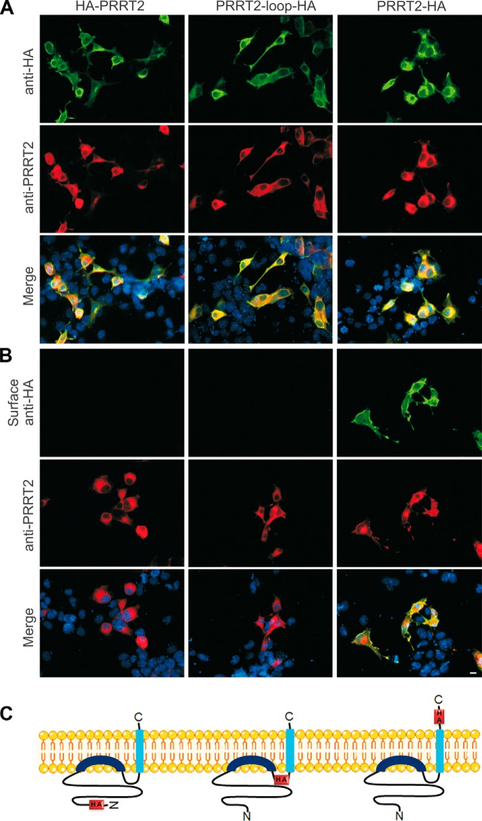
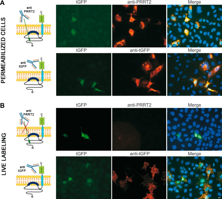
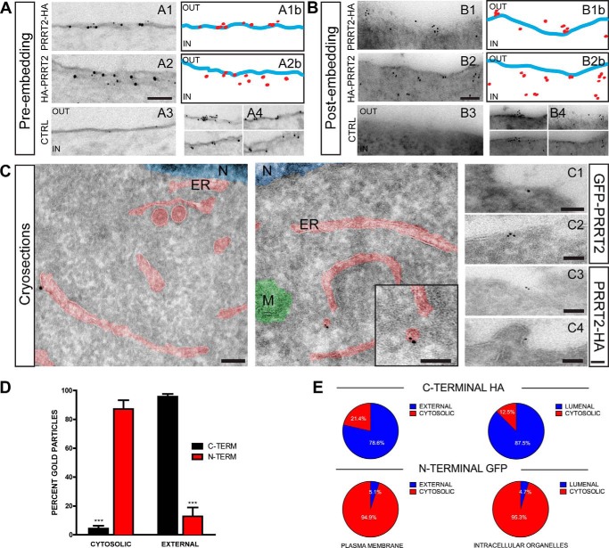
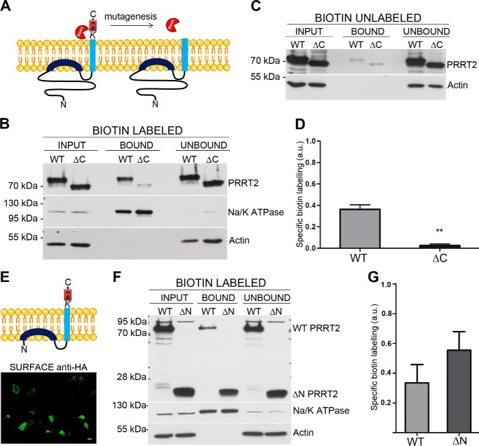
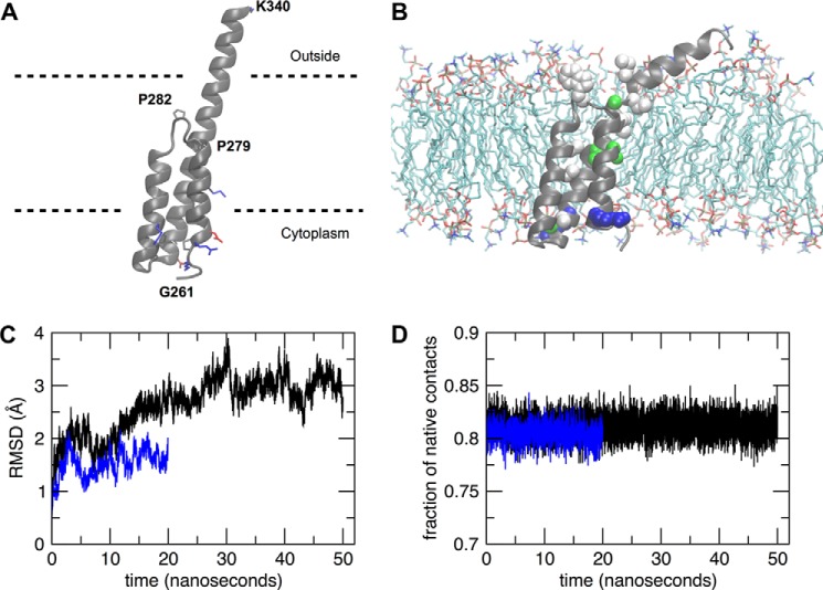
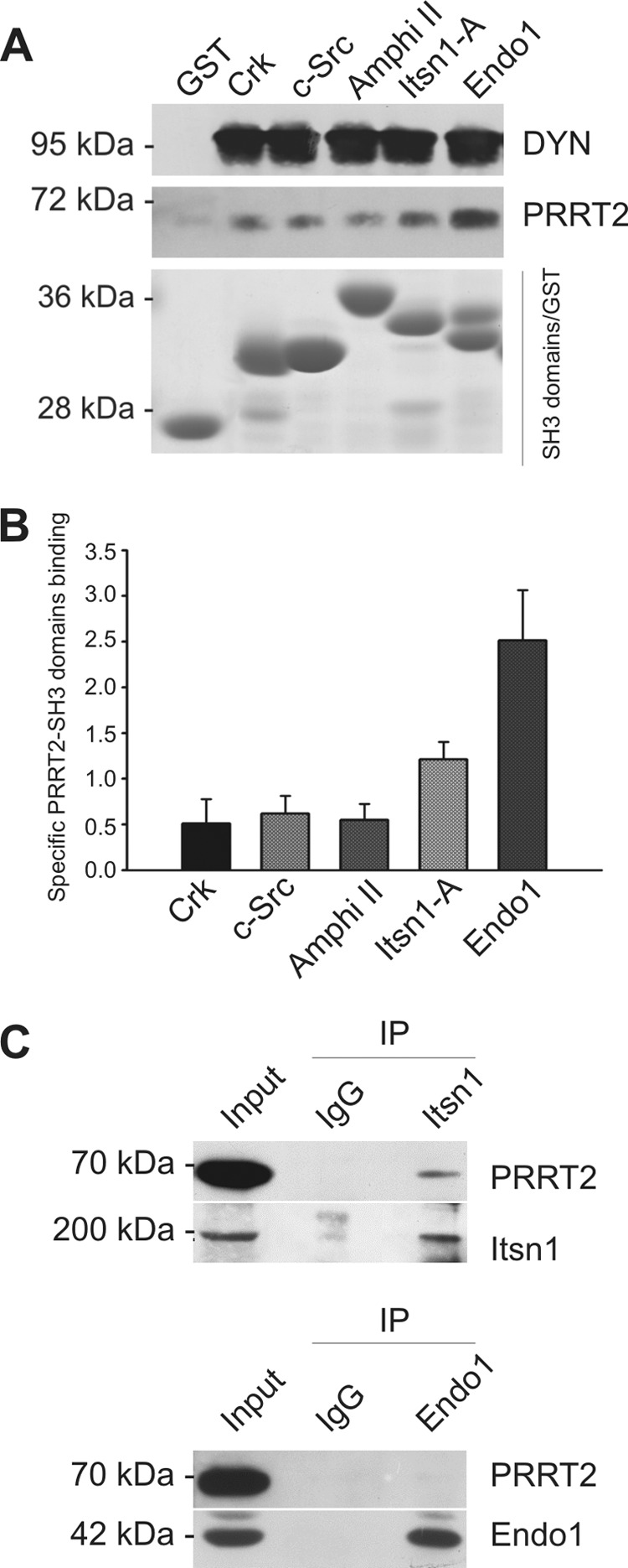
Proline-rich transmembrane protein 2 (PRRT2) has been identified as the single causative gene for a group of paroxysmal syndromes of infancy, including epilepsy, paroxysmal movement disorders, and migraine. On the basis of topology predictions, PRRT2 has been assigned to the recently characterized family of Dispanins, whose members share the two-transmembrane domain topology with a large N terminus and short C terminus oriented toward the outside of the cell. Because PRRT2 plays a role at the synapse, it is important to confirm the exact orientation of its N and C termini with respect to the plasma membrane to get clues regarding its possible function. Using a combination of different experimental approaches, including live immunolabeling, immunogold electron microscopy, surface biotinylation and computational modeling, we demonstrate a novel topology for this protein. PRRT2 is a type II transmembrane protein in which only the second hydrophobic segment spans the plasma membrane, whereas the first one is associated with the internal surface of the membrane and forms a helix-loop-helix structure without crossing it. Most importantly, the large proline-rich N-terminal domain is not exposed to the extracellular space but is localized intracellularly, and only the short C terminus is extracellular (N cyt/C exo topology). Accordingly, we show that PRRT2 interacts with the Src homology 3 domain-bearing protein Intersectin 1, an intracellular protein involved in synaptic vesicle cycling. These findings will contribute to the clarification of the role of PRRT2 at the synapse and the understanding of pathogenic mechanisms on the basis of PRRT2-related neurological disorders.

Keywords: SNARE proteins; membrane protein; molecular dynamics; neurological disease; synapse.


Figures

FIGURE 1.
FIGURE 1.
Different localization of HA epitopes tagging the N and C termini of PRRT2 with respect to the plasma membrane. A, COS7 cells were transfected with distinct HA-tagged PRRT2 constructs (HA-PRRT2, with HA3 tags at the N terminus; PRRT2-loop-HA, with HA3 tags between the two hydrophobic segments in the predicted cytosolic domain; and PRRT2-HA, with HA3 tags at the C terminus), fixed, and permeabilized for standard immunofluorescence. After permeabilization, both anti-HA (top panels) and anti-PRRT2 (center panels) antibodies recognized the overexpressed protein to the same extent (Merge, bottom panels). Cell nuclei were counterstained with DAPI (blue). B, live labeling with anti-HA antibodies only recognized PRRT2-HA, and no signal was detected in cells transfected with PRRT2-loop-HA or HA-PRRT2, indicating that the HA epitopes of these constructs were not accessible on the external surface of the cells (top panels). The different HA constructs were expressed equally, as shown by immunostaining with anti PRRT2 antibodies obtained after permeabilization of the cells (center panels). Bottom panels, merged images from the top and center panels. Scale bar = 20 μm. C, schematic of the membrane topology of PRRT2. The orientation of the N and C termini of PRRT2 with respect to the plasma membrane is shown, with the position of the HA3 tags in the protein sequence of the three constructs used to determine the topology of the protein.
FIGURE 2.
FIGURE 2.
Tagging PRRT2 with an alternative fluorescent epitope confirms the opposite location of the N and C termini with respect to the plasma membrane. A, COS7 cells were transfected with a plasmid encoding for PRRT2 with C-terminal tGFP. After permeabilization, both anti-PRRT2 antibodies recognizing the N terminus (sequence 152–268) and anti-tGFP antibodies detected the transfected protein. B, live labeling with the same antibodies was present only with anti-tGFP antibodies, suggesting that the N-terminal epitopes are not exposed on the external side of the plasma membrane. Scale bar = 20 μm. The different accessibility of PRRT2 epitopes to the antibodies used in each experiment is shown in the left panels. When cells are permeabilized, anti-PRRT2 antibodies can enter the cells and recognize intracellular N-terminal epitopes. Under live labeling condition, the intact plasma membrane blocks their accessibility to N-terminal epitopes.
FIGURE 3.
FIGURE 3.
Ultrastructural localization of the C and N termini of PRRT2 expressed in COS7 cells. A and B, representative micrographs, obtained with the pre-embedding (A) or post-embedding (B) technique, of COS7 cells transfected with either C-terminal PRRT2-HA (A1 and B1) or N-terminal HA-PRRT2 (A2 and B2) and immunogold-labeled with anti-HA antibodies. Non-transfected cells (CTRL, A3 and B3) were also processed as negative controls. A1b and A2b, ImageJ masks of A1 and A2, highlighting the plasma membrane (blue) and the gold particles (red) mapping the N and C termini of PRRT2. B1b and B2b, ImageJ masks of B1 and B2, highlighting the plasma membrane (blue) and the gold particles (red). A4 and B4, additional images of plasma membrane domains of transfected COS7 cells mapping the C-terminal (top panels) or N-terminal PRRT2 (bottom panels). C, representative EM micrographs of PRRT2-expressing COS7 cells obtained with the Tokuyasu cryo-immunogold technique. Cells were co-transfected with N-terminally labeled GFP-PRRT2 and C-terminally labeled PRRT2-HA or left untransfected as a negative control. Representative cryosections labeled with anti-GFP antibodies coupled to 10-nm nanogold particles (left panel) and anti-HA antibodies coupled with 6-nm gold particles (center panel and inset) are shown. Additional images visualizing the location of GFP (C1 and C2) and HA (C3 and C4) immunoreactivities are also shown. N, nucleus; ER, endoplasmic reticulum; M, mitochondrion. Scale bars = 100 nm. D, quantification of the percentage of gold particles located inside (cytosolic) or outside of (external) the plasma membrane from pre- and post-embedding specimens. Error bars represent the mean ± S.E. (n = 46 images from 3 independent experiments) for the C-terminal (black) and N-terminal (red) labeling of PRRT2, respectively. ***, p < 0.01; Student's t test. E, the occurrence of gold particles decorating the C terminus (C-terminal HA) or the N terminus (N-terminal GFP) of PRRT2 calculated from the cryo-immunolabeled sections shown in C. The position of the gold particles was quantified with respect to both the plasma membrane (cytosolic/external) and the membrane of intracellular organelles (cytosolic/lumenal).
FIGURE 4.
FIGURE 4.
PRRT2 is surface-biotinylated at the C-terminal domain. A, biotin labeling of the C-terminal lysine residue of PRRT2 and the lack of labeling of the deletion mutant PRRT2ΔC lacking the C-terminal domain and the HA tag. B, Western blotting analyses probed with anti-PRRT2 antibodies showed biotin labeling of wild-type PRRT2-HA and a strong decrease in biotinylation of PRRT2ΔC. Both isoforms were expressed equally in the cells (input). Their presence in the unbound fraction corresponds to the intracellular pool of the overexpressed protein. Na+/K+ ATPase and actin were used as positive and negative controls of surface biotinylation, respectively. C, representative control experiment in which biotin was omitted. The faint PRRT2 bands represent a low degree of nonspecific binding to the NeutrAvidin beads. D, immunoblots were quantified by densitometric analysis of the fluorograms obtained in the linear range of the emulsion response. The specific biotin labeling of PRRT2 was calculated as [total biotinylation/input − nonspecific biotinylation/input] and is expressed in arbitrary units (a. u.). Specific biotinylation, shown as mean ± S.E. of three independent experiments, is decreased significantly in PRRT2ΔC compared with WT PRRT2-HA. **, p < 0.01; Student's t test for paired samples. E, the PRRT2 mutant lacking the N-terminal domain (PRRT2ΔN) and its plasma membrane expression by live labeling with anti HA antibodies. F and G, representative Western blotting analysis probed with anti-HA antibodies (F) and quantification of the specific biotinylation (G, mean ± S.E., n = 3) showing that both WT PRRT2-HA and PRRT2ΔN are biotinylated specifically to approximately the same extent.
FIGURE 5.
FIGURE 5.
Molecular dynamics simulations of PRRT2 topology. A, predicted model of the PRRT2 protein transmembrane domains (residues 261–340). The protein backbone is shown as a schematic, whereas charged residues are shown as sticks and colored according to their type (blue, basic; red, acidic). Proline residues are also shown as sticks. Dashed lines indicate putative membrane limits. B, snapshot of the protein and membrane system at the end of the 20-ns MD trajectory. Water molecules and some lipids were removed for clarity. The residues mutated in the PRRT2 sequence (6) are shown as spheres and colored according to their type (blue/red, charged; green, polar; white, hydrophobic). C, root mean square displacement (RMSD) of the protein structure with respect to the starting conformation along the 20-ns (blue) and 50-ns (black) MD trajectories. D, fraction of native contacts along the 20-ns (blue) and 50-ns (black) MD trajectories.
FIGURE 6.
FIGURE 6.
Affinity purification of PRRT2 by SH3 domains from extracts of brain synaptosomes A, pulldown assay. Affinity resins were prepared by coupling the SH3 domains of Crk, c-Src, Amphiphysin II (Amphi II), Intersectin 1 SH3-A (Itsn1-A), or Endophilin 1 (Endo1) or GST alone to glutathione-Sepharose. Columns were loaded with a Triton X-100 extract of crude synaptosomal fraction (P2) from rat forebrain. Bound proteins were eluted with SDS, separated by SDS-PAGE, and analyzed by immunoblotting with anti-PRRT2- or anti-dynamin (DYN)-specific antibodies. Center panel, the elution patterns of PRRT2. The immunoreactivity of dynamin I (top panel) is shown for comparison. Bottom panel, recovery of the GST-tagged SH3 domains after Coomassie staining of the gels. B, quantification of the pulldown. Immunoblots were analyzed by densitometry of the fluorograms obtained in the linear range of the emulsion response to quantify band immunoreactivity. The amount of PRRT2 associated with the respective SH3 domains was calculated as the ratio of specific PRRT2-SH3 domain binding (immunoreactivity of the PRRT2 SH3 GST domain − immunoreactivity of PRRT2 GST) with the unspecific binding (immunoreactivity of PRRT2 GST) and expressed as mean ± S.E. (n = 5). C, co-immunoprecipitation. Detergent extracts of mouse brain were immunoprecipitated with anti-intersectin 1 monoclonal antibodies, anti-endophilin-1 monoclonal antibodies, or mouse IgG, as indicated (IP). After electrophoretic separation of the immunocomplexes and Western blotting, membranes were probed with antibodies against Itsn1 and Endo1 to test the efficiency of the immunoprecipitation (bottom blots) and with anti-PRRT2 antibodies (top blots).

References

    1. Méneret A., Gaudebout C., Riant F., Vidailhet M., Depienne C., and Roze E. (2013) PRRT2 mutations and paroxysmal disorders. Eur. J. Neurol. 20, 872–878 - PubMed
    1. Heron S. E., and Dibbens L. M. (2013) Role of PRRT2 in common paroxysmal neurological disorders: a gene with remarkable pleiotropy. J. Med. Genet. 50, 133–139 - PubMed
    1. Becker F., Schubert J., Striano P., Anttonen A. K., Liukkonen E., Gaily E., Gerloff C., Müller S., Heußinger N., Kellinghaus C., Robbiano A., Polvi A., Zittel S., von Oertzen T. J., Rostasy K., Schöls L., Warner T., Münchau A., Lehesjoki A. E., Zara F., Lerche H., and Weber Y. G. (2013) PRRT2-related disorders: further PKD and ICCA cases and review of the literature. J. Neurol. 260, 1234–1244 - PubMed
    1. Guerrini R., and Mink J. W. (2012) Paroxysmal disorders associated with PRRT2 mutations shake up expectations on ion channel genes. Neurology 79, 2086–2088 - PubMed
    1. Wood H. (2012) Genetics: Expanding the spectrum of neurological disorders associated with PRRT2 mutations. Nat. Rev. Neurol. 8, 657. - PubMed

Publication types