. 2016 Mar 25;291(13):6641-54.
doi: 10.1074/jbc.M115.693523. Epub 2016 Jan 21.

Oxidative Stress Promotes Peroxiredoxin Hyperoxidation and Attenuates Pro-survival Signaling in Aging Chondrocytes

Affiliations

Oxidative Stress Promotes Peroxiredoxin Hyperoxidation and Attenuates Pro-survival Signaling in Aging Chondrocytes

John A Collins et al. J Biol Chem. .

Abstract

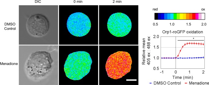
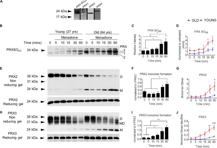
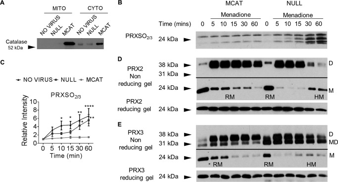
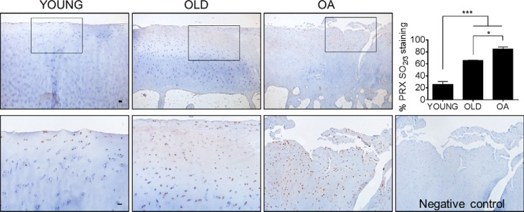
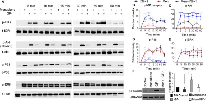
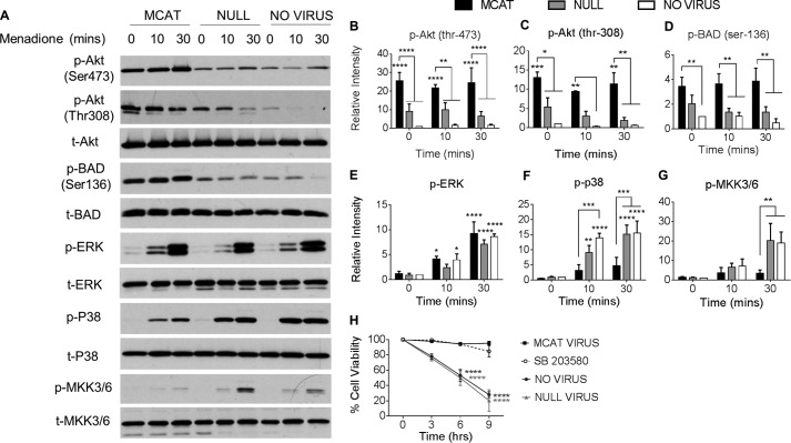
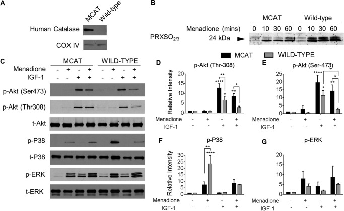
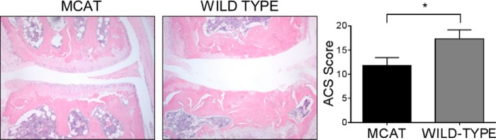
Oxidative stress-mediated post-translational modifications of redox-sensitive proteins are postulated as a key mechanism underlying age-related cellular dysfunction and disease progression. Peroxiredoxins (PRX) are critical intracellular antioxidants that also regulate redox signaling events. Age-related osteoarthritis is a common form of arthritis that has been associated with mitochondrial dysfunction and oxidative stress. The objective of this study was to determine the effect of aging and oxidative stress on chondrocyte intracellular signaling, with a specific focus on oxidation of cytosolic PRX2 and mitochondrial PRX3. Menadione was used as a model to induce cellular oxidative stress. Compared with chondrocytes isolated from young adult humans, chondrocytes from older adults exhibited higher levels of PRX1-3 hyperoxidation basally and under conditions of oxidative stress. Peroxiredoxin hyperoxidation was associated with inhibition of pro-survival Akt signaling and stimulation of pro-death p38 signaling. These changes were prevented in cultured human chondrocytes by adenoviral expression of catalase targeted to the mitochondria (MCAT) and in cartilage explants from MCAT transgenic mice. Peroxiredoxin hyperoxidation was observedin situin human cartilage sections from older adults and in osteoarthritic cartilage. MCAT transgenic mice exhibited less age-related osteoarthritis. These findings demonstrate that age-related oxidative stress can disrupt normal physiological signaling and contribute to osteoarthritis and suggest peroxiredoxin hyperoxidation as a potential mechanism.

Keywords: aging; cell signaling; osteoarthritis; oxidative stress; peroxiredoxin; redox signaling.


Figures

FIGURE 1.
FIGURE 1.
Generation of intracellular H2O2 in human articular chondrocytes treated with menadione. Human articular chondrocytes were transduced with a baculovirus that expresses the H2O2 redox sensor Orp1-roGFP. The cells were treated with or without 25 μm menadione, and the images were collected and analyzed as detailed in the methods. Images from one representative cell for control and menadione-treated conditions are shown in heat map format. Scale bar, 5 μm. Quantified data from 10 individual cells were taken from three independent donors. The data are presented as means ± S.E. showing the relative mean 405:488 ratio. Asterisks represent significant differences comparing menadione-treated chondrocytes to DMSO control. *, p < 0.05 (t test). DIC, differential interference contrast.
FIGURE 2.
FIGURE 2.
Effect of menadione-induced oxidative stress and age on PRX oxidation in human articular chondrocytes. Confluent human articular chondrocytes cultured in serum-free medium were treated for the indicated times with 25 μm menadione to induce oxidative stress. After treatments, cell lysates were prepared in lysis buffer containing 20 mm iodoacetamide to alkylate reduced thiols at lysis. A, human chondrocytes were treated with menadione for 60 min. The sample was loaded onto four lanes of a 12% gel. Each lane was separated by molecular weight marker, and the gel was run under reducing conditions. After protein transfer to membranes, individual lanes were cut out and probed with antibodies to PRXSO2/3, PRX1, PRX2, or PRX3 to confirm the band location of each protein. Comparative analysis confirmed the locations of PRX1–3. B, hyperoxidized PRXs from older and younger chondrocytes were detected using an antibody that reacts with PRX 1–4 when it is in the PRX-SO2/3 state. Specific PRXs are labeled. C, densitometric analysis from immunoblots of n = 6 donors, independent of age. The data are normalized to time 0 untreated controls. D, quantification of PRXSO2/3, normalized to untreated controls from young donors, comparing chondrocytes from older (n = 3; average age, 63 ± 12 years; range, 50–75 years) and younger (n = 3; average age, 38 ± 10 years; range, 27–48 years) donors. E, immunoblots probed for PRX2 under nonreducing conditions (top panel) showing oxidized dimers (labeled D on blots) and hyperoxidized monomers (labeled M on blots). Immunoblotting for PRX2 under reducing conditions was used as a loading control (lower panel). F, densitometric analysis of PRX2 hyperoxidized monomer from n = 6 independent donors, independent of age. G, quantification of the PRX2 monomer:dimer ratio from the same donors as in D. H, immunoblots probed for PRX3 under nonreducing conditions (top panel). Hyperoxidized monomer is labeled M on blots, mixed dimers are labeled MD on blots, and oxidized dimer is labeled D on blots. Immunoblotting for PRX3 under reducing conditions was used as a loading control (lower panel). I, densitometric analysis of PRX3 hyperoxidized monomer from n = 6 independent donors, independent of age. J, quantification of the PRX3 monomer:mixed disulfide ratio from same donors as in D. The data are presented as means ± S.E. Asterisks represent significant differences between younger and older chondrocytes. *, p < 0.05; **, p < 0.01; ***, p < 0.0001) (ANOVA). yrs, years.
FIGURE 3.
FIGURE 3.
The effects of serum-free culture and cell lysis conditions on PRX oxidation. A, confluent human chondrocytes were either cultured in the presence of serum (10%) or serum-starved for 16 h, and basal levels of reduced, oxidized, and hyperoxidized PRX2 and PRX3 were detected by immunoblot. Monomeric PRX is labeled M on blots, and dimeric bands are labeled D on blots. PRX3 mixed disulfides are labeled MD on blots. B, densitometric analysis from immunoblots of n = 3 donors, independent of age. The data are normalized to β-actin loading control. The data are presented as means ± S.E. NS denotes nonsignificant differences between treatments. C, PRX hyperoxidation in human chondrocytes treated with IAM at lysis or NEM pretreatment (10 min) prior to NEM at lysis was detected with the antibody to PRX-SO2/3. D and E, under nonreducing conditions, immunoblots for total PRX2 and PRX3 allowed for direct comparison of the IAM and NEM pretreatment methods to detect the redox status of PRXs. Monomeric bands are labeled M on blots, reduced monomers are labeled RM on blots, and hyperoxidized monomers are labeled HM on blots. PRX3 presented with an oxidized dimer (labeled D on blots), as well as a lower mixed disulfide band (labeled MD on blots). Solid lines on blots are used to separate different exposures from the same immunoblot.
FIGURE 4.
FIGURE 4.
Targeted expression of human catalase to the mitochondria attenuates menadione-induced PRX oxidation in human chondrocytes. Confluent human articular chondrocytes were transduced with an adenovirus expressing mitochondrial catalase (MCAT), a null (empty) vector control, or no virus for 48 h prior to overnight incubation in serum-free medium and addition of 25 μm menadione for the indicated times to induce oxidative stress. Chondrocyte monolayers were incubated in 100 mm NEM for 10 min to alkylate reduced thiols prior to lysis, and cell lysates were prepared in NEM containing lysis buffer prior to immunoblotting. A, immunoblot of chondrocyte mitochondrial and cytosolic fractions showing expression of human catalase protein expression in unstimulated chondrocytes transduced with MCAT adenovirus, a null empty vector, or no virus. B, chondrocyte cell lysates transduced with MCAT adenovirus, a null empty vector, or no virus and then treated with menadione for the indicated times. Hyperoxidized PRXs were identified using the antibody to PRXSO2/3. C, results of densitometric analysis from PRXSO2/3 immunoblots from n = 3 independent donors. D, under nonreducing conditions, immunoblots for total PRX2 allowed for identification of the PRX2 reduced monomer (labeled RM on blots) and the hyperoxidized monomer (labeled HM on blots). PRX2 presented with an oxidized dimer (labeled D on blots). E, under nonreducing conditions, immunoblots for total PRX3 allowed for identification of the PRX3 reduced monomer (labeled RM on blots) and the hyperoxidized monomer (labeled HM on blots). PRX3 presented with an oxidized dimer (labeled D on blots), as well as a lower mixed disulfide band (labeled MD on blots). Solid lines on blots are used to separate different exposures from the same immunoblot. All immunoblots are representative results of n = 3 donors. The data are means ± S.E. normalized to untreated controls. Asterisks represent significant differences compared with control. *, p < 0.05; **, p < 0.01; ****, p < 0.0001 (ANOVA).
FIGURE 5.
FIGURE 5.
Hyperoxidized PRX is observed in human cartilage from older adults and in osteoarthritic cartilage. The levels of PRXSO2/3 in normal younger, normal older, and osteoarthritic human knee cartilage was evaluated by immunohistochemistry using the antibody to PRXSO2/3. Tissue sections were counterstained with hematoxylin. Human cartilage sections (n = 4 independent samples per group) were macroscopically graded as described under “Experimental Procedures.” For these experiments, younger donors were <50 years old (mean age, 41.3 ± 6.1 years), and older donors were ≥50 years old (mean age, 73.3 ± 4.3 years). The average age of the OA patients was 79.3 ± 13.6 years. Scale bar, 100 μm. Representative negative control consists of primary incubation in antibody diluent without addition of anti-PRXSO2/3. The data are presented as means ± S.E. of percentage PRXSO2/3 stained cells for each condition. Asterisks represent significant differences compared with control. *, p < 0.05; ***, p < 0.001 (ANOVA).
FIGURE 6.
FIGURE 6.
Effect of oxidative stress on IGF-1 and MAP kinase signaling in normal human chondrocytes. Confluent human articular chondrocytes cultured in serum-free medium were treated for the indicated times (0–90 min) with menadione (25 μm) or IGF-1 (50 ng/ml). For combined treatments, chondrocytes were pretreated with menadione (25 μm) for 30 min prior to IGF-1 treatment for the indicated times (0–90 min). After experimental incubations, cell lysates were prepared and immunoblotted under reducing conditions with antibodies to phosphorylated IGF receptor, Akt, ERK, and p38. The blots were then stripped and reprobed with respective total antibodies for loading controls. A, representative immunoblots from three independent experiments. B–E, results of densitometric analysis showing phosphorylation of IGF receptor, Akt, ERK, and p38. F, chondrocytes were treated for 30 min with 25 μm menadione, treated 30 min with 50 ng/ml IGF-1, or pretreated with menadione (30 min) prior to IGF-1 treatment (30 min), and immunoblotting was performed on reducing gels to probe for phosphorylation of PRAS40, a downstream marker of Akt activity. Phosphorylation of proteins was normalized to total protein as a loading control and presented as relative change compared with untreated controls. The data are presented as means ± S.E. from n = 3 donors. Asterisks represent significant differences compared with menadione treatment at that time point. *, p < 0.05; **, p < 0.01; ***, p < 0.001; ****, p < 0.0001 (ANOVA). MEN, menadione.
FIGURE 7.
FIGURE 7.
Expression of mitochondrial-targeted catalase increases Akt phosphorylation and attenuates pro-apoptotic signaling induced by oxidative stress. Confluent human articular chondrocytes were transduced with an adenovirus expressing mitochondrial catalase (MCAT), a null (empty) vector control, or no virus for 48 h prior to overnight incubation in serum-free medium and addition of 25 μm menadione for the indicated times (0–30 min) to induce oxidative stress. After experimental incubations, cell lysates were prepared and immunoblotted under reducing conditions with antibodies to phosphorylated Akt, BAD, ERK, p38, and MKK3/6. The blots were then stripped and reprobed with respective total antibodies for loading controls. A, representative immunoblots from three independent experiments. B–G, results of densitometric analysis showing phosphorylation of Akt, BAD, ERK, p38, and MKK3/6. Phosphorylation of proteins were normalized to total protein as a loading control and presented as fold change compared with untreated controls. The data are presented as means ± S.E. from n = 3 independent donors. H, chondrocytes treated with virus as above were treated with 25 μm menadione for the indicated times (0–9 h) to induce oxidative stress. For p38 inhibitor experiments, chondrocytes were treated with the p38 inhibitor SB203580 (10 μm) for 30 min prior to menadione treatment. Cell survival was assessed as described under “Experimental Procedures.” The data are quantified as percentages of cell death (means ± S.E.) compared with control values from a minimum of three independent experiments. Asterisks represent significant differences compared with no virus untreated controls. *, p < 0.05; **, p < 0.01; ***, p < 0.001; ****, p < 0.0001 (ANOVA).
FIGURE 8.
FIGURE 8.
Effect of oxidative stress on PRX hyperoxidation and kinase phosphorylation in chondrocytes from MCAT and wild-type mice. Femoral head cartilage explants from the hip joints of 4-week-old MCAT and wild-type mice were isolated and cultured in serum-supplemented medium for 48 h prior to overnight incubation in serum-free medium. Femoral head cartilage explants were treated with menadione (25 μm), IGF-1 (50 ng/ml), or combined treatments as indicated. Cell lysates were prepared by homogenization of explants as described under “Experimental Procedures.” A, cell lysates were immunoblotted for human catalase and for COX IV as a mitochondrial protein loading control. B, femoral cap cartilage explants from MCAT and wild-type mice were cultured in menadione for 0–60 min prior to incubation in NEM (100 mm, 10 min) to alkylate reduced thiols prior to lysis. Femoral caps were disrupted and homogenized in lysis buffer containing NEM. Hyperoxidized PRXs were detected by immunoblot using the PRX-SO2/3 antibody. C, for analysis of signaling, femoral cap cartilage explants were treated with menadione (30 min), IGF-1 (30 min), or pretreated with menadione (30 min) prior to IGF-1 treatment (30 min). Representative blots from n = 3 independent experiments are shown. D–G, results of densitometric analysis showing phosphorylation of Akt, p38, and ERK. Phosphorylation of proteins were normalized to total protein as a loading control and presented as fold change from untreated control. All data are presented as means ± S.E. from n = 3 independent experiments. Asterisks represent significant differences compared with control. *, p < 0.05; **, p < 0.01; ***, p < 0.001; ****, p < 0.0001 (ANOVA).
FIGURE 9.
FIGURE 9.
MCAT mice develop less severe age-related OA. Histological evaluation of knee joints from MCAT and wild-type mice aged 18–33 months. Sections from age-matched wild-type and MCAT mice are shown. Independent blinded histological assessment of OA severity was conducted on the medial and lateral tibial plateaus using an OA grading scheme (Articular Cartilage Structure score). The data are presented as means ± S.E. of the medial plus lateral tibial plateau Articular Cartilage Structure Scores n = 5 wild-type and n = 6 MCAT mice. The asterisk represents a significant difference between mouse phenotypes. *, p < 0.05 (ANOVA).

Similar articles

Cited by

References

    1. López-Otín C., Blasco M. A., Partridge L., Serrano M., and Kroemer G. (2013) The hallmarks of aging. Cell 153, 1194–1217 - PMC - PubMed
    1. van Deursen J. M. (2014) The role of senescent cells in ageing. Nature 509, 439–446 - PMC - PubMed
    1. Balaban R. S., Nemoto S., and Finkel T. (2005) Mitochondria, oxidants, and aging. Cell 120, 483–495 - PubMed
    1. Chan D. C. (2006) Mitochondria: dynamic organelles in disease, aging, and development. Cell 125, 1241–1252 - PubMed
    1. Finkel T. (2011) Signal transduction by reactive oxygen species. J. Cell Biol. 194, 7–15 - PMC - PubMed

Publication types

MeSH terms