

doi: 10.1021/acschembio.5b00900.
Epub 2016 Mar 1.
Five-Membered Ring Peroxide Selectively Initiates Ferroptosis in Cancer Cells
Affiliations
- PMID: 26797166
- PMCID: PMC5507670
- DOI: 10.1021/acschembio.5b00900
Item in Clipboard
Five-Membered Ring Peroxide Selectively Initiates Ferroptosis in Cancer Cells
ACS Chem Biol.
.
Abstract
A 1,2-dioxolane (FINO2) was identified as a lead compound from a screen of organic peroxides. FINO2 does not induce apoptosis, but instead initiates ferroptosis, an iron-dependent, oxidative cell death pathway. Few compounds are known to induce primarily ferroptosis. In contrast to the perceived instability of peroxides, FINO2 was found to be thermally stable to at least 150 °C. FINO2 was more potent in cancer cells than nonmalignant cells of the same type. One of the enantiomers was found to be more responsible for the observed activity.
Conflict of interest statement
The authors declare no competing financial interest.
Figures
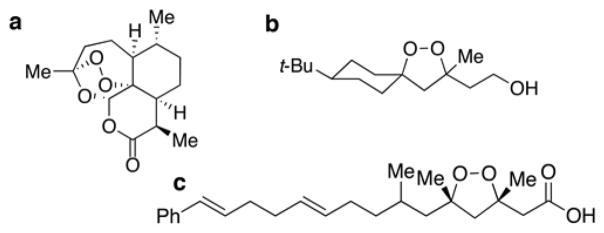
Chemical structures of peroxides. (a) Artemisinin. (b) Lead 1,2-dioxolane (FINO2). (c) Plakinic acid D.

Comparison of FINO2 to FDA-approved anticancer agents. (a) GI50, TGI (total growth inhibition), and LC50 values for FINO2 compared to three other anticancer agents. (b) Average (AVG), standard deviation (σ), and coefficient of variation (CV) of FDA-approved chemotherapeutic agents compared to FINO2 to demonstrate similarity in the amount of variation between cell lines.
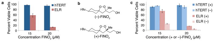
Selectivity of FINO2 measured by Promega CellTiter–Glo Luminescence Assay. (a) Comparison of noncancerous (BJ-hTERT; blue) and oncogenically transformed (BJ-ELR; red) fibroblast cells treated with the racemic form of FINO2. (b) Structures of enantiomers of FINO2. (c) Comparison of noncancerous (BJ-hTERT; blue) and oncogenically transformed (BJ-ELR; red) cells treated with individual enantiomers of FINO2. All experiments were performed in triplicate, and error bars reflect the standard deviation of one experiment. A representative graph of at least three independent experiments is shown.

A nonapoptotic form of cell death induced by FINO2. (a) Annexin V staining is not observed prior to 7AAD incorporation with 6 μM FINO2 treatment at indicated time points. (b) RS4;11 cells overexpressing either GFP or Bcl-2 treated with and FINO2 (left) and etoposide (right). Percent dead cells indicate the Annexin V and 7AAD positive cell population. The nature of the steep dose–response curve induced by FINO2 causes a slight difference in concentrations necessary to induce cell death depending on the preparation of the stock solutions of FINO2 and the passage number of the cells. All experiments were performed at a large concentration range, and the appropriate data are reported.

Many features of ferroptosis exhibited by FINO2-induced cell death. (a) Electron microscopy image of typical RS4;11 cell treated with FINO2 showing a lack of hallmarks of apoptosis, necrosis, and autophagy (performed by the Microscopy Core at New York University Langone Medical Center). Detection of oxidative stress 6 h after indicated treatment by (b) CellROX Green Reagent, Molecular Probes, and (c) BODIPY 581/591 C11. (d) Dependence on iron is shown by iron chelation with 20 μM deferoxamine (DFO) or iron addition with 38 μM ferric ammonium citrate (FAC). (e) Pretreatment with lipophilic antioxidants 500 nM ferrostatin and (f) 200 nM liproxstatin prevent cell death. (g) Pretreatment with 5 μM nordihydroguaiaretic acid (NDGA) prevents cell death. (h) Pretreatment with 100 μM indomethacin partially inhibits cell death. Percent dead cells indicate the Annexin V and 7AAD positive cell population. The nature of the steep dose–response curve induced by FINO2 causes a slight difference in concentrations necessary to induce cell death depending on the preparation of the stock solutions of FINO2 and the passage number of the cells. All experiments were performed at a large concentration range, and the appropriate data are reported.
References
-
- Siegel R, Naishadham D, Jemal A. Cancer Statistics, 2013. Ca-Cancer J Clin. 2013;63:11–30. - PubMed
-
- Siegel RL, Miller KD, Jemal A. Cancer Statistics, 2015. Ca-Cancer J Clin. 2015;65:5–29. - PubMed
-
- Holohan C, Van Schaeybroeck S, Longley DB, Johnston PG. Cancer drug resistance: an evolving paradigm. Nat Rev Cancer. 2013;13:714–726. - PubMed
-
- Chonghaile TN, Sarosiek KA, Vo TT, Ryan JA, Tammareddi A, Moore VDG, Deng J, Anderson KC, Richardson P, Tai YT, Mitsiades CS, Matulonis UA, Drapkin R, Stone R, DeAngelo DJ, McConkey DJ, Sallan SE, Silverman L, Hirsch MS, Carrasco DR, Letai A. Pretreatment Mitochondrial Priming Correlates with Clinical Response to Cytotoxic Chemotherapy. Science. 2011;334:1129–1133. - PMC - PubMed
-
- Czabotar PE, Lessene G, Strasser A, Adams JM. Control of apoptosis by the BCL-2 protein family: implications for physiology and therapy. Nat Rev Mol Cell Biol. 2014;15:49–63. - PubMed
Publication types
MeSH terms
Substances
Grants and funding
LinkOut - more resources
Full Text Sources
Other Literature Sources
