

Analysis of free drug fractions in serum by ultrafast affinity extraction and two-dimensional affinity chromatography using α1-acid glycoprotein microcolumns
- PMID: 26797422
- PMCID: PMC4724389
- DOI: 10.1016/j.chroma.2015.12.084
Analysis of free drug fractions in serum by ultrafast affinity extraction and two-dimensional affinity chromatography using α1-acid glycoprotein microcolumns
Abstract
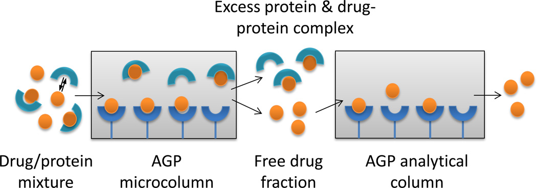
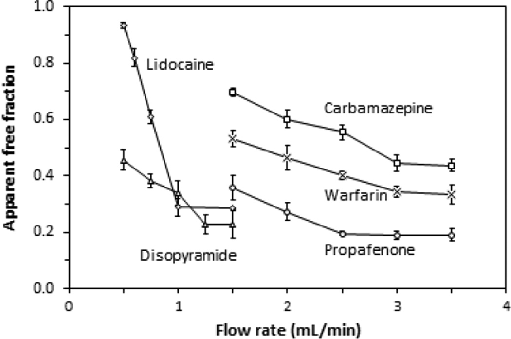
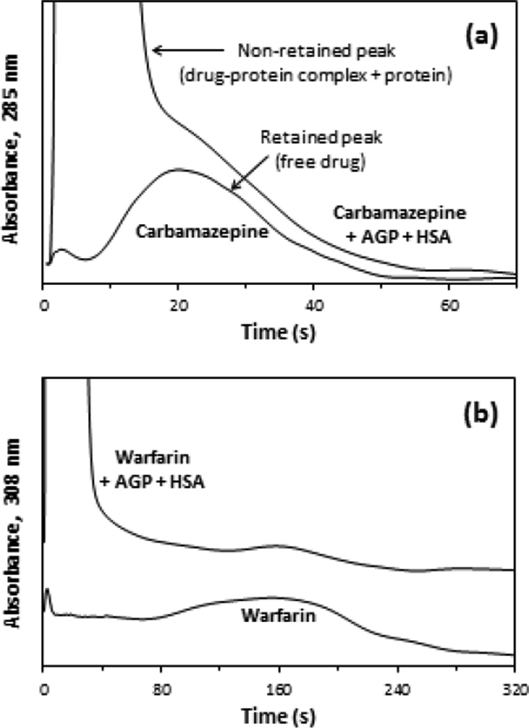
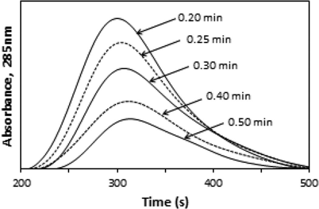
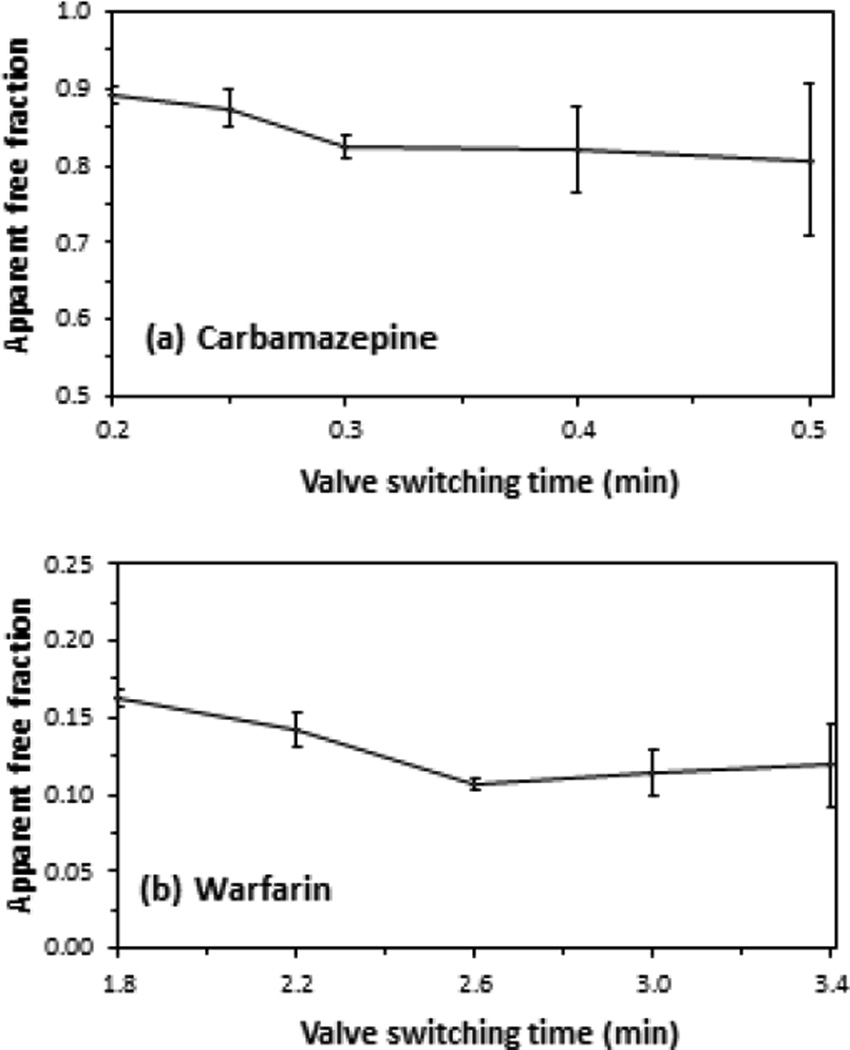
In the circulatory system, many drugs are reversibly bound to serum proteins such as human serum albumin (HSA) and alpha1-acid glycoprotein (AGP), resulting in both free and protein-bound fractions for these drugs. This report examined the use of microcolumns containing immobilized AGP for the measurement of free drug fractions by ultrafast affinity extraction and a two-dimensional affinity system. Several drugs known to bind AGP were used as models to develop and evaluate this approach. Factors considered during the creation of this method included the retention of the drugs on the microcolumns, the injection flow rate, the microcolumn size, and the times at which a second AGP column was placed on-line with the microcolumn. The final system had residence times of only 110-830ms during sample passage through the AGP microcolumns and allowed free drug fractions to be determined within 10-20min when using only 3-10μL of sample per injection. This method was used to measure the free fractions of the model drugs at typical therapeutic levels in serum, giving good agreement with the results obtained by ultrafiltration. This approach was also used to estimate the binding constants for each drug with AGP in serum, even for drugs that had significant interactions with both AGP and HSA in such samples. These results indicated that AGP microcolumns could be used with ultrafast affinity extraction to measure free drug fractions in a label-free manner and to study the binding of drugs with AGP in complex samples such as serum.
Keywords: Drug-protein binding; Free drug fraction; Human serum albumin; Ultrafast affinity extraction; α(1)-Acid glycoprotein.
Copyright © 2015 Elsevier B.V. All rights reserved.
Figures
Similar articles
-
Characterization of drug binding with alpha1-acid glycoprotein in clinical samples using ultrafast affinity extraction.J Chromatogr A. 2021 Jul 19;1649:462240. doi: 10.1016/j.chroma.2021.462240. Epub 2021 May 11. J Chromatogr A. 2021. PMID: 34034105 Free PMC article.
-
Analysis of free drug fractions in human serum by ultrafast affinity extraction and two-dimensional affinity chromatography.Anal Bioanal Chem. 2016 Jan;408(1):131-40. doi: 10.1007/s00216-015-9082-7. Epub 2015 Oct 13. Anal Bioanal Chem. 2016. PMID: 26462924 Free PMC article.
-
Analysis of free drug fractions by ultrafast affinity extraction: interactions of sulfonylurea drugs with normal or glycated human serum albumin.J Chromatogr A. 2014 Dec 5;1371:82-9. doi: 10.1016/j.chroma.2014.10.092. Epub 2014 Oct 31. J Chromatogr A. 2014. PMID: 25456590 Free PMC article.
-
[Study on binding of drug to serum protein].Yakugaku Zasshi. 2009 Apr;129(4):413-25. doi: 10.1248/yakushi.129.413. Yakugaku Zasshi. 2009. PMID: 19336995 Review. Japanese.
-
A molecular functional study on the interactions of drugs with plasma proteins.Drug Metab Pharmacokinet. 2005 Oct;20(5):309-23. doi: 10.2133/dmpk.20.309. Drug Metab Pharmacokinet. 2005. PMID: 16272748 Review.
Cited by
-
Analysis of the binding of warfarin to glyoxal- and methylglyoxal-modified human serum albumin by ultrafast affinity extraction.J Chromatogr B Analyt Technol Biomed Life Sci. 2022 Nov 15;1211:123500. doi: 10.1016/j.jchromb.2022.123500. Epub 2022 Oct 13. J Chromatogr B Analyt Technol Biomed Life Sci. 2022. PMID: 36272357 Free PMC article.
-
High performance affinity chromatography and related separation methods for the analysis of biological and pharmaceutical agents.Analyst. 2018 Jan 15;143(2):374-391. doi: 10.1039/c7an01469d. Analyst. 2018. PMID: 29200216 Free PMC article. Review.
-
Affinity chromatography: A review of trends and developments over the past 50 years.J Chromatogr B Analyt Technol Biomed Life Sci. 2020 Nov 10;1157:122332. doi: 10.1016/j.jchromb.2020.122332. Epub 2020 Aug 14. J Chromatogr B Analyt Technol Biomed Life Sci. 2020. PMID: 32871378 Free PMC article. Review.
-
[Advances in chromatography in the study of drug-plasma protein interactions].Se Pu. 2021 Oct;39(10):1077-1085. doi: 10.3724/SP.J.1123.2021.06028. Se Pu. 2021. PMID: 34505429 Free PMC article. Review. Chinese.
-
Evaluation of microcolumn stability in ultrafast affinity extraction for binding and rate studies.J Chromatogr B Analyt Technol Biomed Life Sci. 2021 Dec 15;1187:123047. doi: 10.1016/j.jchromb.2021.123047. Epub 2021 Nov 17. J Chromatogr B Analyt Technol Biomed Life Sci. 2021. PMID: 34823097 Free PMC article.
References
-
- Vuignier K, Schappler J, Veuthey JL, Carrupt PA, Martel S. Drug-protein binding: a critical review of analytical tools. Anal. Bioanal. Chem. 2010;398:53–66. - PubMed
Publication types
MeSH terms
Substances
Grants and funding
LinkOut - more resources
Full Text Sources
Other Literature Sources
Medical
