. 2016 Jan 22;351(6271):379-84.
doi: 10.1126/science.aad3839.

Oligodendrocyte precursors migrate along vasculature in the developing nervous system

Affiliations

Oligodendrocyte precursors migrate along vasculature in the developing nervous system

Hui-Hsin Tsai et al. Science. .

Abstract

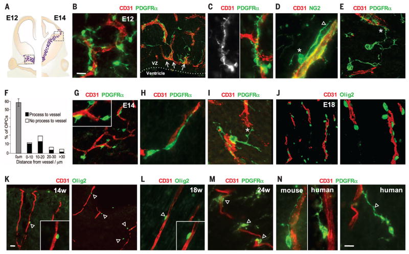
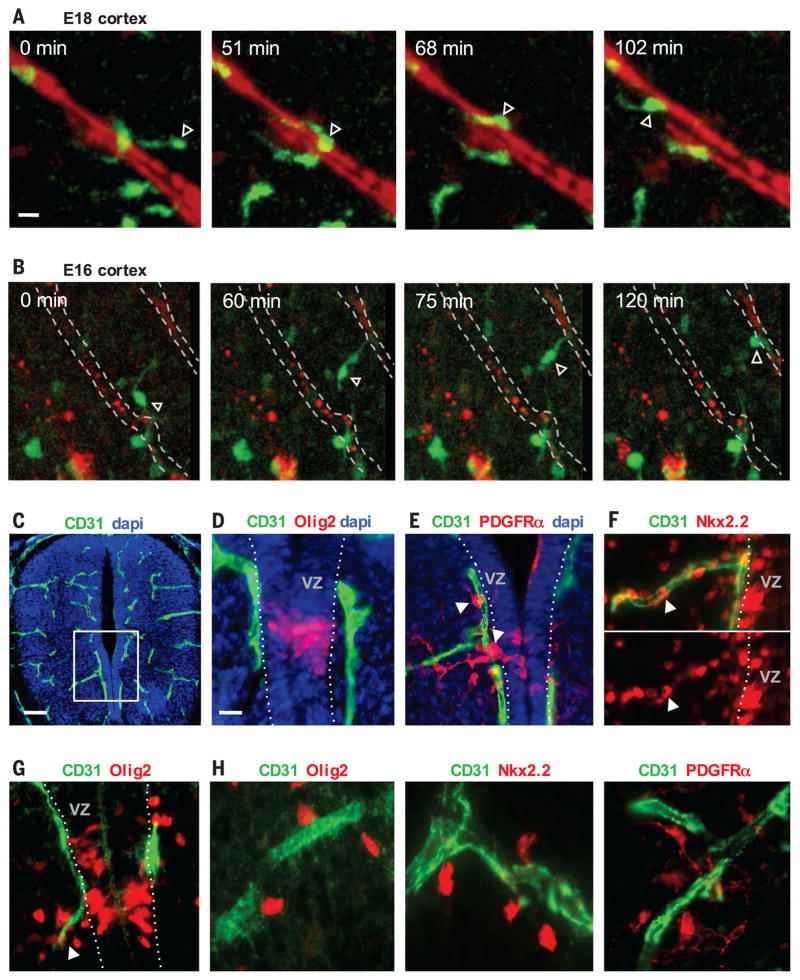
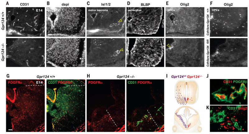
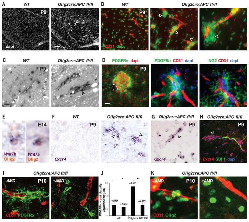
Oligodendrocytes myelinate axons in the central nervous system and develop from oligodendrocyte precursor cells (OPCs) that must first migrate extensively during brain and spinal cord development. We show that OPCs require the vasculature as a physical substrate for migration. We observed that OPCs of the embryonic mouse brain and spinal cord, as well as the human cortex, emerge from progenitor domains and associate with the abluminal endothelial surface of nearby blood vessels. Migrating OPCs crawl along and jump between vessels. OPC migration in vivo was disrupted in mice with defective vascular architecture but was normal in mice lacking pericytes. Thus, physical interactions with the vascular endothelium are required for OPC migration. We identify Wnt-Cxcr4 (chemokine receptor 4) signaling in regulation of OPC-endothelial interactions and propose that this signaling coordinates OPC migration with differentiation.


Figures

Fig. 1
Fig. 1. Association of migratory OPCs with vessels in the developing mouse and human brain
(A) OPCs (dots) first appear from the ventral medial ganglionic eminence of the mouse brain at E12 (left) and migrate away from the ventricular zone to reach the subpallial-to-pallial boundary at E14 (right). (B to E) Images taken from the boxed area on the left side of (A). (B) PDGFRα+ migratory OPCs at E12 associate tightly with CD31+ vasculature and show elongated morphology along vessels (C), often with cell bodies on the endothelial surface [* in (D)], extending a leading process along [arrowhead in (D)] or toward [* in (E)] a vessel. VZ, ventricular zone. (F) Quantification of OPC association with vessels at E12 (1056 cells measured, n = 4 animals). Error bar indicates SD. (G to I) Images taken from the boxed area on the right side of (A). Migratory OPCs at the subpallial-to-pallial boundary at E14 maintain tight association with the vasculature (G), with process extension along (H) and between [* in (I)] vessels. Inset in (G) shows zoomed-in view. (J) Migrating Olig2+ OPCs in the E18 cortex palisade along cortical penetrating vessels. (K to N) (K) The first OPCs (Olig2+ and PDGFRα+) to arrive in the developing human outer cortex closely associate with vessels at gestational weeks 14 (14w) (K), 18 (L), and 24 (M) and show similar morphological association with vessels as in mice [left, (N)], with leading processes that can be as long as 65 μm [arrowhead, right (N)]. Scale bars: 10 μm [(B) and (N)]; 20 μm (K).
Fig. 2
Fig. 2. OPC crawling and jumping directed motility on vessels
(A and B) Time-lapse imaging in slice cultures of the embryonic Olig2-GFP mouse cortex showing GFP expression (green) and vessels (red). (A) A GFP-expressing OPC (arrowhead) crawls along a penetrating vessel in the E18 cortex (movie S1). (B) An OPC in the E16 cortex (arrowhead) (movie S3) demonstrates jumping motility between vessels, extending a process to a parallel cortical penetrating vessel, before movement of the cell body onto the new vessel. (C and D) Olig2-expressing OPCs define the pMN domain of the spinal cord ventricular zone at E12. (D) shows a zoomed-in view of the boxed area in (C). dapi, 4′,6-diamidino-2-phenylindole. (E and F) The first PDGFRα- and Nkx2.2-expressing OPCs (arrowheads) emerge from the pMN immediately onto and along the adjacent CD31+ vasculature. (G and H) (G) OPCs tend to leave the pMN at E14 along a vessel that branches at the pMN (arrowhead), and these cells remain associated with vessels as they continue to disperse throughout the ventral spinal cord (H). Scale bars: 10 μm [(A) and (D)]; 40 μm (C).
Fig. 3
Fig. 3. OPCs require a vascular scaffold for migration
(A and B) (A) Gpr124−/− embryos exhibit CNS vascular patterning defects at E14, leaving the ventral spinal cord [dapi in (B)] with reduced vascularization compared with controls. (C) Islet1/2+ motor neurons migrate in normal numbers at E14, from the pMN to ventrolateral gray matter (arrowheads) in Gpr124−/− cords. (D) BLBP-expressing astrocytes also migrate normally from ventricular zone domains adjacent to the pMN into the surrounding gray matter in Gpr124−/−. (E and F) (E) OPC (Olig2+) emigration from the ventricular zone is severely disrupted in Gpr124−/− [(E) and (I)] and Cdh5cre/Gpr124-floxed (F) spinal cord with accumulation of OPCs in the pMN [arrowhead in (E)]. (G) By E14, OPCs (PDGFRα+) in wild-type (WT) Gpr124+/+ brains migrate as far as the subpallial-to-pallial boundary (dotted line). (H and I) (H) Vascular development (CD31+ vessels) is highly truncated in the brains of E14 Gpr124−/− mice, with associated severe OPC migration deficits (I). OPCs migrate only as far as the limits of the ventral vascular plexus [dotted line in (H)]. (J and K) (J) Absence of Gpr124 leads to vascular abnormalities (glomeruloid malformations) in which OPCs (Olig2+) accumulate in clumps in the E14 Gpr124−/− brain (K). Scale bars: 30 μm [(A) and (B)]; 60 μm (G).
Fig. 4
Fig. 4. A Wnt-activated, Cxcr4-dependent mechanism drives OPC attraction to the vasculature
(A to D) (A) Aberrant accumulations of cells (arrowheads) appear in the P9 corpus callosum associated with the CD31+ vasculature (B) of Olig2-cre:Apcflox/flox mice; can be seen in resin sections stained with Toluidine blue (C); and represent OPCs, as labeled by PDGFRα and NG2 staining (D). (E) OPCs produce Wnt7a and Wnt7b at E14 during embryonic migration. (F to H) (F) Wnt activation in P9 Olig2-cre:Apcflox/flox corpus callosum (F) or spinal cord (G) leads to marked up-regulation (compared with wild type) of Cxcr4 mRNA [(F) and (G)] and protein (H) in clustered OPCs (arrowheads) associated with SDF1-expressing endothelium (H). (I) Treatment of Olig2-cre:Apcflox/flox spinal cord with the Cxcr4/SDF1 inhibitor AMD3100 (+AMD) leads to a reduction in clustered OPCs associated with the vasculature, as compared with controls (−AMD). (J) Number of PDGFRα+ OPCs on a vessel in P10 spinal cord in WT versus untreated or AMD3100-treated Olig2-cre:Apcflox/flox mice, expressed as number of cells on each 100-μm CD31 vessel segment (*P = 8.9 × 10⁻⁵, **P = 1.9 × 10⁻⁴, Bonferroni test, n = 4 animals per group). (K) Treatment of P10 corpus callosum Olig2-cre:Apcflox/flox slices ex vivo overnight with 10 μg/ml of AMD3100 (+AMD, right panel) leads to a reduction in vessel-associated OPC clustering, as compared with untreated controls (−AMD, left panel). Scale bars: 120 μm (A); 10 μm [(C) and (D)].

Comment in

References

    1. Fünfschilling U, et al. Nature. 2012;485:517–521. - PMC - PubMed
    1. Lee Y, et al. Nature. 2012;487:443–448. - PMC - PubMed
    1. Lu QR, et al. Cell. 2002;109:75–86. - PubMed
    1. Kessaris N, et al. Nat Neurosci. 2006;9:173–179. - PMC - PubMed
    1. Sidman RL, Rakic P. Brain Res. 1973;62:1–35. - PubMed

Publication types

MeSH terms