Coronary Microembolization with Normal Epicardial Coronary Arteries and No Visible Infarcts on Nitrobluetetrazolium Chloride-Stained Specimens: Evaluation with Cardiac Magnetic Resonance Imaging in a Swine Model
- PMID: 26798220
- PMCID: PMC4720817
- DOI: 10.3348/kjr.2016.17.1.83
Coronary Microembolization with Normal Epicardial Coronary Arteries and No Visible Infarcts on Nitrobluetetrazolium Chloride-Stained Specimens: Evaluation with Cardiac Magnetic Resonance Imaging in a Swine Model
Abstract
Objective: To assess magnetic resonance imaging (MRI) features of coronary microembolization in a swine model induced by small-sized microemboli, which may cause microinfarcts invisible to the naked eye.
Materials and methods: Eleven pigs underwent intracoronary injection of small-sized microspheres (42 µm) and catheter coronary angiography was obtained before and after microembolization. Cardiac MRI and measurement of cardiac troponin T (cTnT) were performed at baseline, 6 hours, and 1 week after microembolization. Postmortem evaluation was performed after completion of the imaging studies.
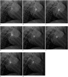
Results: Coronary angiography pre- and post-microembolization revealed normal epicardial coronary arteries. Systolic wall thickening of the microembolized regions decreased significantly from 42.6 ± 2.0% at baseline to 20.3 ± 2.3% at 6 hours and 31.5 ± 2.1% at 1 week after coronary microembolization (p < 0.001 for both). First-pass perfusion defect was visualized at 6 hours but the extent was largely decreased at 1 week. Delayed contrast enhancement MRI (DE-MRI) demonstrated hyperenhancement within the target area at 6 hours but not at 1 week. The microinfarcts on gross specimen stained with nitrobluetetrazolium chloride were invisible to the naked eye and only detectable microscopically. Increased cTnT was observed at 6 hours and 1 week after microembolization.
Conclusion: Coronary microembolization induced by a certain load of small-sized microemboli may result in microinfarcts invisible to the naked eye with normal epicardial coronary arteries. MRI features of myocardial impairment secondary to such microembolization include the decline in left ventricular function and myocardial perfusion at cine and first-pass perfusion imaging, and transient hyperenhancement at DE-MRI.
Keywords: Cardiac MR imaging; Coronary angiography; Myocardial contraction; Myocardial microinfarct.
Figures


Similar articles
-
Assessment of the acute effects of glucocorticoid treatment on coronary microembolization using cine, first-pass perfusion, and delayed enhancement MRI.J Magn Reson Imaging. 2016 Apr;43(4):921-8. doi: 10.1002/jmri.25049. Epub 2015 Sep 11. J Magn Reson Imaging. 2016. PMID: 26361889
-
Acute hyperenhancement on delayed contrast-enhanced magnetic resonance imaging is the characteristic sign after coronary microembolization.Chin Med J (Engl). 2009 Mar 20;122(6):687-91. Chin Med J (Engl). 2009. PMID: 19323935
-
Coronary microembolization causes long-term detrimental effects on regional left ventricular function.Scand Cardiovasc J. 2011 Aug;45(4):205-14. doi: 10.3109/14017431.2011.568629. Epub 2011 Apr 4. Scand Cardiovasc J. 2011. PMID: 21463182
-
Coronary microembolization--its role in acute coronary syndromes and interventions.Herz. 1999 Nov;24(7):558-75. doi: 10.1007/BF03044228. Herz. 1999. PMID: 10609163 Review.
-
[Magnetic resonance tomography imaging techniques for diagnosing myocardial vitality].Herz. 1994 Feb;19(1):51-64. Herz. 1994. PMID: 8150414 Review. German.
Cited by
-
Decreased atrioventricular plane displacement after acute myocardial infarction yields a concomitant decrease in stroke volume.J Appl Physiol (1985). 2020 Feb 1;128(2):252-263. doi: 10.1152/japplphysiol.00480.2019. Epub 2019 Dec 19. J Appl Physiol (1985). 2020. PMID: 31854250 Free PMC article.
References
-
- Porto I, Selvanayagam JB, Van Gaal WJ, Prati F, Cheng A, Channon K, et al. Plaque volume and occurrence and location of periprocedural myocardial necrosis after percutaneous coronary intervention: insights from delayed-enhancement magnetic resonance imaging, thrombolysis in myocardial infarction myocardial perfusion grade analysis, and intravascular ultrasound. Circulation. 2006;114:662–669. - PubMed
-
- Baumgart D, Liu F, Haude M, Görge G, Ge J, Erbel R. Acute plaque rupture and myocardial stunning in patient with normal coronary arteriography. Lancet. 1995;346:193–194. - PubMed
-
- Falk E. Unstable angina with fatal outcome: dynamic coronary thrombosis leading to infarction and/or sudden death. Autopsy evidence of recurrent mural thrombosis with peripheral embolization culminating in total vascular occlusion. Circulation. 1985;71:699–708. - PubMed
-
- Kotani J, Nanto S, Mintz GS, Kitakaze M, Ohara T, Morozumi T, et al. Plaque gruel of atheromatous coronary lesion may contribute to the no-reflow phenomenon in patients with acute coronary syndrome. Circulation. 2002;106:1672–1677. - PubMed
-
- Henriques JP, Zijlstra F, Ottervanger JP, de Boer MJ, van't Hof AW, Hoorntje JC, et al. Incidence and clinical significance of distal embolization during primary angioplasty for acute myocardial infarction. Eur Heart J. 2002;23:1112–1117. - PubMed
Publication types
MeSH terms
Substances
LinkOut - more resources
Full Text Sources
Other Literature Sources
Medical
Research Materials

