

Concordant and Discordant Cardiac Magnetic Resonance Imaging Delayed Hyperenhancement Patterns in Patients with Ischemic and Non-Ischemic Cardiomyopathy
- PMID: 26798384
- PMCID: PMC4720848
- DOI: 10.4070/kcj.2016.46.1.41
Concordant and Discordant Cardiac Magnetic Resonance Imaging Delayed Hyperenhancement Patterns in Patients with Ischemic and Non-Ischemic Cardiomyopathy
Abstract
Background and objectives: The diagnosis of ischemic (ICM) and non-ischemic cardiomyopathy (NICM) is conventionally determined by the presence or absence of coronary artery disease (CAD) in the setting of a reduced left systolic function. However the presence of CAD may not always indicate that the actual left ventricular (LV) dysfunction mechanism is ischemia, as other non-ischemic etiologies can be responsible. We investigated patterns of myocardial fibrosis using delayed hyperenhancement (DHE) on cardiac magnetic resonance (CMR) in ICM and NICM.
Subjects and methods: Patients with systolic heart failure who underwent a CMR were prospectively analyzed. The heart failure diagnosis was based on the modified Framingham criteria and LVEF <35%. LV dysfunction was classified as ICM or NICM based on coronary anatomy.
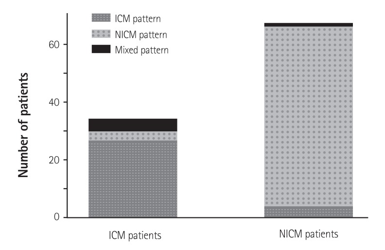
Results: A total of 101 subjects were analyzed; 34 were classified as ICM and 67 as NICM. The DHE pattern was concordant with the conventional diagnosis in 27 (79.4%) of the patients with ICM and 62 (92.5%) of the patients with NCIM. A discordant NICM DHE pattern was present in 8.8% of patients with ICM, and an ICM pattern was detected 6.0% of the patients with NICM. Furthermore, 11.8% of the patients with ICM and 1.5% of those with NICM demonstrated a mixed pattern.
Conclusion: A subset of patients conventionally diagnosed with ICM or NICM based on coronary anatomy demonstrated a discordant or mixed DHE pattern. CMR-DHE imaging can be helpful to determine the etiology of heart failure in patients with persistent LV systolic dysfunction.
Keywords: Cardiac imaging technique; Cardiomyopathies; Magnetic resonance imaging.
Conflict of interest statement
The authors have no financial conflicts of interest.
Figures
Similar articles
-
Nonischemic or Dual Cardiomyopathy in Patients With Coronary Artery Disease.Circulation. 2024 Mar 12;149(11):807-821. doi: 10.1161/CIRCULATIONAHA.123.067032. Epub 2023 Nov 6. Circulation. 2024. PMID: 37929565 Free PMC article.
-
Late gadolinium enhancement from cardiac magnetic resonance in ischemic and non-ischemic cardiomyopathy.J Med Assoc Thai. 2011 Feb;94 Suppl 1:S33-8. J Med Assoc Thai. 2011. PMID: 21721426
-
Differentiating Nonischemic Dilated Cardiomyopathy With Incidental Infarction From Ischemic Cardiomyopathy by Geometric Indices Derived From Cardiovascular Magnetic Resonance.J Thorac Imaging. 2021 Jul 1;36(4):248-253. doi: 10.1097/RTI.0000000000000560. J Thorac Imaging. 2021. PMID: 32960835
-
Myocardial Fibrosis Assessment by LGE Is a Powerful Predictor of Ventricular Tachyarrhythmias in Ischemic and Nonischemic LV Dysfunction: A Meta-Analysis.JACC Cardiovasc Imaging. 2016 Sep;9(9):1046-1055. doi: 10.1016/j.jcmg.2016.01.033. Epub 2016 Jul 20. JACC Cardiovasc Imaging. 2016. PMID: 27450871 Review.
-
Differences in the Clinical Outcome of Ischemic and Nonischemic Cardiomyopathy in Heart Failure With Concomitant Opioid Use Disorder.Curr Probl Cardiol. 2023 May;48(5):101609. doi: 10.1016/j.cpcardiol.2023.101609. Epub 2023 Jan 21. Curr Probl Cardiol. 2023. PMID: 36690309 Review.
Cited by
-
A Novel Approach for Identifying Ischemic Cardiomyopathy.Korean Circ J. 2016 Jan;46(1):13-4. doi: 10.4070/kcj.2016.46.1.13. Epub 2016 Jan 14. Korean Circ J. 2016. PMID: 26798380 Free PMC article. No abstract available.
-
Practical management of peripartum cardiomyopathy.Korean J Intern Med. 2017 May;32(3):393-403. doi: 10.3904/kjim.2016.360. Epub 2017 Apr 14. Korean J Intern Med. 2017. PMID: 28407464 Free PMC article. Review.
-
Echocardiography-based machine learning algorithm for distinguishing ischemic cardiomyopathy from dilated cardiomyopathy.BMC Cardiovasc Disord. 2023 Sep 26;23(1):476. doi: 10.1186/s12872-023-03520-4. BMC Cardiovasc Disord. 2023. PMID: 37752424 Free PMC article.
-
Nonischemic or Dual Cardiomyopathy in Patients With Coronary Artery Disease.Circulation. 2024 Mar 12;149(11):807-821. doi: 10.1161/CIRCULATIONAHA.123.067032. Epub 2023 Nov 6. Circulation. 2024. PMID: 37929565 Free PMC article.
References
-
- Nieminen MS, Böhm M, Cowie MR, et al. Executive summary of the guidelines on the diagnosis and treatment of acute heart failure: the Task Force on Acute Heart Failure of the European Society of Cardiology. Eur Heart J. 2005;26:384–416. - PubMed
-
- McCrohon JA, Moon JC, Prasad SK, et al. Differentiation of heart failure related to dilated cardiomyopathy and coronary artery disease using gadolinium-enhanced cardiovascular magnetic resonance. Circulation. 2003;108:54–59. - PubMed
-
- Wu E, Judd RM, Vargas JD, Klocke FJ, Bonow RO, Kim RJ. Visualisation of presence, location, and transmural extent of healed Q-wave and non-Q-wave myocardial infarction. Lancet. 2001;357:21–28. - PubMed
LinkOut - more resources
Full Text Sources
Other Literature Sources
Miscellaneous
