. 2016:2016:2075915.
doi: 10.1155/2016/2075915. Epub 2015 Dec 21.

t-BHQ Provides Protection against Lead Neurotoxicity via Nrf2/HO-1 Pathway

Affiliations

t-BHQ Provides Protection against Lead Neurotoxicity via Nrf2/HO-1 Pathway

Fang Ye et al. Oxid Med Cell Longev. 2016.

Abstract

The neurotoxicity of lead has been well established, and oxidative stress is strongly associated with lead-induced neurotoxicity. Nrf2 is important for protection against oxidative stress in many disease models. We applied t-BHQ, which is an Nrf2 activator, to investigate the possible role of Nrf2 in the protection against lead neurotoxicity. t-BHQ significantly attenuated the oxidative stress in developmental rats by decreasing MDA level, as well as by increasing SOD activity and GSH content, in the hippocampus and frontal cortex. Furthermore, neuronal apoptosis was detected by Nissl staining, and Bax expression was inhibited in the t-BHQ-treated group. Results showed that t-BHQ suppressed ROS production and caspase 3/7 activity but increased intracellular GSH content, in SH-SY5Y cells under lead exposure. Moreover, in vivo and in vitro, t-BHQ enhanced the nuclear translocation of Nrf2 and binding to ARE areas but did not induce Nrf2 transcription. These phenomena were confirmed using RT-PCR, EMSA, Western blot, and immunofluorescence analyses. Subsequent upregulation of the expression of HO-1, NQO1, and GCLC was observed. However, knockdown of Nrf2 or HO-1 adversely affected the protective effects of t-BHQ against lead toxicity in SH-SY5Y cells. Thus, t-BHQ can protect against lead neurotoxicity, depending on the Nrf2/HO-1 pathway.


Figures
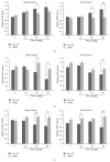
Figure 1
Figure 1
t-BHQ alleviates PbAc-induced alterations in redox status in the hippocampus and frontal cortex. Rats pretreated with t-BHQ (150 mg/kg) or corn oil for three days by oral gavage then cotreated with varies doses (10, 30 or 60 mg/kg) PbAc for five days by intraperitoneal injection (ip). The levels of MDA (a), SOD activity (b), and GSH content (c) were detected in these eight groups in the hippocampus and frontal cortex. n = 5 in each group. Data represent the mean ± SEM of the results. P < 0.05 and ∗∗ P < 0.01 represent significant differences compared with the control group (corn oil without PbAc). # P < 0.05 and ## P < 0.01 represent significant differences between two groups with or without t-BHQ treatment at the same concentration of PbAc.
Figure 2
Figure 2
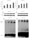
t-BHQ promotes Nrf2 nuclear translocation but does not induce Nrf2 transcription in the hippocampus and frontal cortex. (a) Nrf2 mRNA level was quantified by RT-PCR and the appropriate dose of PbAc (60 mg/kg) was chosen in this experiment. n = 5 in each group. (b and c) Nuclear protein was isolated and applied in Western blot experiment (b) and EMSA experiment (c). Nrf2-ARE binding activity and the content of Nrf2 in nucleus were detected and the immunoblots were quantified. n = 5 in each group. Data represent the mean ± SEM of the results. P < 0.05 represents significant differences compared with the control group.
Figure 3
Figure 3
t-BHQ upregulates the expression of Nrf2 downstream targets in the hippocampus and frontal cortex. Rats were pretreated with t-BHQ or corn oil and then subjected to PbAc exposure (60 mg/kg). The levels of mRNAs and protein were examined by RT-PCR and Western blot, respectively. The mRNA of GCLC, HO-1, and NQO1 (a) and the protein levels of HO-1 and NQO1 (b) were analyzed in both the hippocampus and frontal cortex. n = 5 in each group. Data represent the mean ± SEM of the results. P < 0.05, ∗∗ P < 0.01, and ∗∗∗ P < 0.001 represent significant differences compared with the control group. # P < 0.05 and ## P < 0.01 represent significant differences between two groups with or without t-BHQ treatment at the same concentration of PbAc (60 mg/kg).
Figure 4
Figure 4
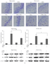
Administration of t-BHQ protects rats against PbAc toxicity. (a) Nissl staining of rat brain sections was applied to investigate the histomorphology of cells in the hippocampus CA1 region and frontal cortex. The injured neurons (arrows) were characterized by cytoplasmic shrinkage, nuclear pyknosis, or hyperchromasia. Scar bar = 50 μm. The ratios of death neuroes/total neuroes were caculated and analyzed in these two regions, respectively. Data were collected from four fields of view. (b) The proapoptotic protein of Bax and antiapoptotic protein of Bcl2 were detected in the hippocampus and frontal cortex; immunoblots were quantified, respectively. n = 5 in each group. Data represent the mean ± SEM of the results. ∗∗ P < 0.01 and ∗∗∗ P < 0.001 represent significant differences compared with the control group. # P < 0.05 and ## P < 0.01 represent significant differences between two groups with or without t-BHQ treatment at the same concentration of PbAc (60 mg/kg).
Figure 5
Figure 5
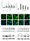
t-BHQ promotes Nrf2 tranlocation from cytoplasm to nucleus and induces the Nrf2-regulated gene expression, especially HO-1. (a) the mRNAs of HO-1, NQO1, GCLC, and Nrf2 were detected by RT-PCR, when cells were treated by t-BHQ (40 μM) for various time (3, 6, 12, or 24 h). (b and c) The immunofluorescent assay was applied to investigate Nrf2 transloction from cytoplasm to nucleus. Scar bar = 20 μm. The content of Nrf2 in nucleus and cytoplasm was analyzed by ImageJ and the ratio of nue/cyt was caculated. n = 3 in each group. (d) Cells were treated by t-BHQ at various doses (10, 20, 40, and 80 μM) or for various time (3, 6, 12, and 24 h); the protein of HO-1 was detected by Western blot. (e) Cells were pretreated with Act.D (0.5 μg/mL) or CHX (10 μg/mL) for 1 h and then treated with t-BHQ or DMSO for 12 h. Cellular proteins were extracted for HO-1 detection. (f) Cells were transfected with 60 pmols control siRNA or Nrf2 siRNA for 24 h and then treated with t-BHQ (40 μM) for an additional 12 h; the HO-1 protein was detected by immunoblot. Data represent the mean ± SEM of the results. P < 0.05, ∗∗ P < 0.01, and ∗∗∗ P < 0.001 represent significant differences compared with the control group.
Figure 6
Figure 6
t-BHQ confers protection against PbAc-induced oxidative stress and apoptosis. The SH-SY5Y cells were pretreated with t-BHQ (40 μM) or DMSO for 12 h, followed by PbAc exposure (25 μM) for 24 h. Cell viability (a), caspase 3/7 activity (b), ROS production (c), GSH content (d), and apoptotic related proteins of Bcl2, Bax, and Bcl-xl (e) were analyzed. All data represent the mean ± SEM of the results. P < 0.05, ∗∗ P < 0.01, and ∗∗∗ P < 0.001 represent significant differences.
Figure 7
Figure 7
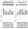
Nrf2/HO-1 pathway mediates the protection of t-BHQ against lead toxicity. (a) and (b) con-siRNA, Nrf2-siRNA, or HO-1-siRNA were transfected into SH-SY5Y cells for 12 h before treatment with t-BHQ. The potein levels of Nrf2 and HO-1 were analyzed by Western blot. (c–f) Cells were transfected with con-siRNA, Nrf2-siRNA, or HO-1-siRNA for 12 h and treated with t-BHQ or DMSO for another 12 h and then exposed to PbAc for 24 h in the presence of t-BHQ or DMSO as indicated. Cell viability (c) and caspase 3/7 activity (d) were measured and analyzed. Western blot analyzed the proapoptotic protein of Bax and antiapoptotic protein of Bcl2 in cells transfected with Nrf2-siRNA (e) or HO-1 siRNA (f) and then treated with t-BHQ (40 μM) and PbAc (25 μM) as indicated above. All data represent the mean ± SEM of the results. P < 0.05 and ∗∗ P < 0.01 represent significant differences.

Similar articles

Cited by

References

    1. White L. D., Cory-Slechta D. A., Gilbert M. E., et al. New and evolving concepts in the neurotoxicology of lead. Toxicology and Applied Pharmacology. 2007;225(1):1–27. doi: 10.1016/j.taap.2007.08.001. - DOI - PubMed
    1. Needleman H. Lead poisoning. Annual Review of Medicine. 2004;55(1):209–222. doi: 10.1146/annurev.med.55.091902.103653. - DOI - PubMed
    1. Lidsky T. I., Schneider J. S. Lead neurotoxicity in children: basic mechanisms and clinical correlates. Brain. 2003;126(1):5–19. doi: 10.1093/brain/awg014. - DOI - PubMed
    1. Yiin S. J., Lin T. H. Lead-catalyzed peroxidation of essential unsaturated fatty acid. Biological Trace Element Research. 1995;50(2):167–172. doi: 10.1007/bf02789419. - DOI - PubMed
    1. Matés J. M. Effects of antioxidant enzymes in the molecular control of reactive oxygen species toxicology. Toxicology. 2000;153(1–3):83–104. doi: 10.1016/s0300-483x(00)00306-1. - DOI - PubMed

Publication types

MeSH terms

LinkOut - more resources