

Mutations in the Parkinson's Disease-Associated PARK2 Gene Are Accompanied by Imbalance in Programmed Cell Death Systems
- PMID: 26798503
- PMCID: PMC4717261
Mutations in the Parkinson's Disease-Associated PARK2 Gene Are Accompanied by Imbalance in Programmed Cell Death Systems
Abstract
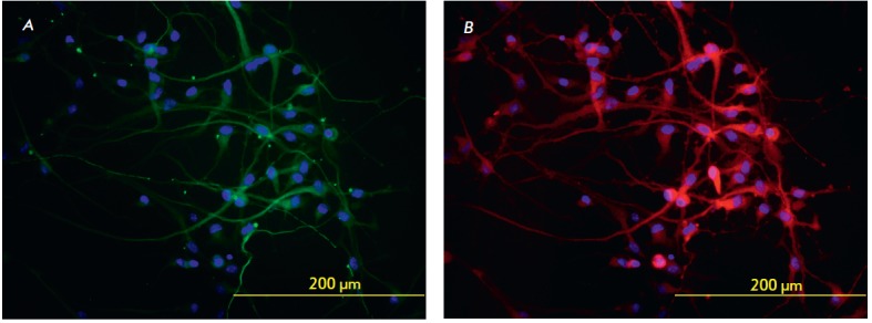
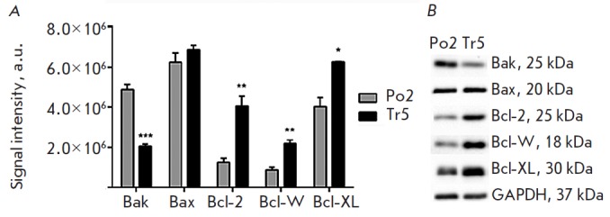
Parkinson's disease is caused by the degeneration of midbrain dopaminergic neurons. A rare recessive form of the disease may be caused by a mutation in the PARK2 gene, whose product, Parkin, controls mitophagy and programmed cell death. The level of pro- and anti-apoptotic factors of the Bcl-2 family was determined in dopaminergic neurons derived from the induced pluripotent stem cells of a healthy donor and a Parkinson's disease patient bearing PARK2 mutations. Western blotting was used to study the ratios of Bax, Bak, Bcl-2, Bcl-XL, and Bcl-W proteins. The pro-apoptotic Bak protein level in PARK2-neurons was shown to be two times lower than that in healthy cells. In contrast, the expression of the anti-apoptotic factors Bcl-XL, Bcl-W, and Bcl-2 was statistically significantly higher in the mutant cells compared to healthy dopaminergic neurons. These results indicate that PARK2 mutations are accompanied by an imbalance in programmed cell death systems in which non-apoptotic molecular mechanisms play the leading role.
Keywords: PARK2; Parkinson’s disease; dopaminergic neurons; induced pluripotent stem cells; mutation; programmed cell death.
Figures
References
-
- Bonifati V.. Parkinsonism Relat. Disord. 2014;20(1):S23–S28. - PubMed
-
- Zagorovskaya T.B., Illarioshkin S.N., Slominskii P.A., Ivanova-Smolenskaya I.A., Markova E.D., Limborskaya S.A., Levin O.S., Miloserdova O.V., Proskokova T.N., Bagyeva G.H., Bris A., S.S. Korsakov zhurn. nevrologii i psikhiatrii. 2004;8:66–72. - PubMed
-
- Kilarski L.L., Pearson J.P., Newsway V., Majounie E., Knipe M.D., Misbahuddin A., Chinnery P.F., Burn D.J., Clarke C.E., Marion M.H.. Mov. Disord. 2012;27:1522–1529. - PubMed
LinkOut - more resources
Full Text Sources
Research Materials