Immunomodulatory effects of amniotic membrane matrix incorporated into collagen scaffolds
- PMID: 26799369
- PMCID: PMC5030101
- DOI: 10.1002/jbm.a.35663
Immunomodulatory effects of amniotic membrane matrix incorporated into collagen scaffolds
Abstract
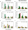
Adult tendon wound repair is characterized by the formation of disorganized collagen matrix which leads to decreases in mechanical properties and scar formation. Studies have linked this scar formation to the inflammatory phase of wound healing. Instructive biomaterials designed for tendon regeneration are often designed to provide both structural and cellular support. In order to facilitate regeneration, success may be found by tempering the body's inflammatory response. This work combines collagen-glycosaminoglycan scaffolds, previously developed for tissue regeneration, with matrix materials (hyaluronic acid and amniotic membrane) that have been shown to promote healing and decreased scar formation in skin studies. The results presented show that scaffolds containing amniotic membrane matrix have significantly increased mechanical properties and that tendon cells within these scaffolds have increased metabolic activity even when the media is supplemented with the pro-inflammatory cytokine interleukin-1 beta. Collagen scaffolds containing hyaluronic acid or amniotic membrane also temper the expression of genes associated with the inflammatory response in normal tendon healing (TNF-α, COLI, MMP-3). These results suggest that alterations to scaffold composition, to include matrix known to decrease scar formation in vivo, can modify the inflammatory response in tenocytes. © 2016 Wiley Periodicals, Inc. J Biomed Mater Res Part A: 104A: 1332-1342, 2016.
Keywords: 3D scaffold; amniotic membrane; collagen; inflammation; tissue engineering.
© 2016 Wiley Periodicals, Inc.
Figures


Similar articles
-
Evaluation of two distinct placental-derived membranes and their effect on tenocyte responses in vitro.J Tissue Eng Regen Med. 2019 Aug;13(8):1316-1330. doi: 10.1002/term.2876. Epub 2019 Jun 13. J Tissue Eng Regen Med. 2019. PMID: 31062484 Free PMC article.
-
Tenocyte cell density, migration, and extracellular matrix deposition with amniotic suspension allograft.J Orthop Res. 2019 Feb;37(2):412-420. doi: 10.1002/jor.24173. Epub 2018 Nov 30. J Orthop Res. 2019. PMID: 30378182 Free PMC article.
-
The effect of cell debris within biologic scaffolds upon the macrophage response.J Biomed Mater Res A. 2017 Aug;105(8):2109-2118. doi: 10.1002/jbm.a.36055. Epub 2017 Apr 12. J Biomed Mater Res A. 2017. PMID: 28263432
-
Human Amniotic Membrane: A review on tissue engineering, application, and storage.J Biomed Mater Res B Appl Biomater. 2021 Aug;109(8):1198-1215. doi: 10.1002/jbm.b.34782. Epub 2020 Dec 14. J Biomed Mater Res B Appl Biomater. 2021. PMID: 33319484 Review.
-
Extracellular Matrix Bioscaffolds as Immunomodulatory Biomaterials<sup/>Tissue Eng Part A. 2017 Oct;23(19-20):1152-1159. doi: 10.1089/ten.TEA.2016.0538. Epub 2017 May 19. Tissue Eng Part A. 2017. PMID: 28457179 Free PMC article. Review.
Cited by
-
Amnion and chorion matrix maintain hMSC osteogenic response and enhance immunomodulatory and angiogenic potential in a mineralized collagen scaffold.Front Bioeng Biotechnol. 2022 Nov 14;10:1034701. doi: 10.3389/fbioe.2022.1034701. eCollection 2022. Front Bioeng Biotechnol. 2022. PMID: 36466348 Free PMC article.
-
Potential application of aminiotic stem cells in veterinary medicine.Anim Reprod. 2020 May 22;16(1):24-30. doi: 10.21451/1984-3143-AR2018-00124. Anim Reprod. 2020. PMID: 33299475 Free PMC article.
-
Mechanome-Guided Strategies in Regenerative Rehabilitation.Curr Opin Biomed Eng. 2024 Mar;29:100516. doi: 10.1016/j.cobme.2023.100516. Epub 2023 Nov 30. Curr Opin Biomed Eng. 2024. PMID: 38586151 Free PMC article.
-
Incorporating β-cyclodextrin into collagen scaffolds to sequester growth factors and modulate mesenchymal stem cell activity.Acta Biomater. 2018 Aug;76:116-125. doi: 10.1016/j.actbio.2018.06.033. Epub 2018 Jun 23. Acta Biomater. 2018. PMID: 29944975 Free PMC article.
-
Application of Amniotic Membrane in Skin Regeneration.Pharmaceutics. 2023 Feb 23;15(3):748. doi: 10.3390/pharmaceutics15030748. Pharmaceutics. 2023. PMID: 36986608 Free PMC article. Review.
References
-
- James R, Kesturu G, Balian G, Chhabra AB. Tendon: Biology, biomechanics, repair, growth factors, and evolving treatment options. J Hand Surg Am. 2008;33:102–112. - PubMed
-
- Lin TW, Cardenas L, Soslowsky LJ. Biomechanics of tendon injury and repair. J Biomech. 2004;37:865–877. - PubMed
-
- Diegelmann RF, Evans MC. Wound healing: an overview of acute, fibrotic and delayed healing. Front Biosci. 2004;9:283–289. - PubMed
-
- Eming SA, Krieg T, Davidson JM. Inflammation in wound repair: molecular and cellular mechanisms. J Invest Dermatol. 2007;127:514–525. - PubMed
Publication types
MeSH terms
Substances
Grants and funding
LinkOut - more resources
Full Text Sources
Other Literature Sources
Miscellaneous

