A New Generation of FRET Sensors for Robust Measurement of Gαi1, Gαi2 and Gαi3 Activation Kinetics in Single Cells
- PMID: 26799488
- PMCID: PMC4723041
- DOI: 10.1371/journal.pone.0146789
A New Generation of FRET Sensors for Robust Measurement of Gαi1, Gαi2 and Gαi3 Activation Kinetics in Single Cells
Abstract
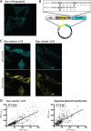
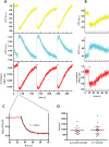
G-protein coupled receptors (GPCRs) can activate a heterotrimeric G-protein complex with subsecond kinetics. Genetically encoded biosensors based on Förster resonance energy transfer (FRET) are ideally suited for the study of such fast signaling events in single living cells. Here we report on the construction and characterization of three FRET biosensors for the measurement of Gαi1, Gαi2 and Gαi3 activation. To enable quantitative long-term imaging of FRET biosensors with high dynamic range, fluorescent proteins with enhanced photophysical properties are required. Therefore, we use the currently brightest and most photostable CFP variant, mTurquoise2, as donor fused to Gαi subunit, and cp173Venus fused to the Gγ2 subunit as acceptor. The Gαi FRET biosensors constructs are expressed together with Gβ1 from a single plasmid, providing preferred relative expression levels with reduced variation in mammalian cells. The Gαi FRET sensors showed a robust response to activation of endogenous or over-expressed alpha-2A-adrenergic receptors, which was inhibited by pertussis toxin. Moreover, we observed activation of the Gαi FRET sensor in single cells upon stimulation of several GPCRs, including the LPA2, M3 and BK2 receptor. Furthermore, we show that the sensors are well suited to extract kinetic parameters from fast measurements in the millisecond time range. This new generation of FRET biosensors for Gαi1, Gαi2 and Gαi3 activation will be valuable for live-cell measurements that probe Gαi activation.
Conflict of interest statement
Figures


Similar articles
-
A FRET-based biosensor for measuring Gα13 activation in single cells.PLoS One. 2018 Mar 5;13(3):e0193705. doi: 10.1371/journal.pone.0193705. eCollection 2018. PLoS One. 2018. PMID: 29505611 Free PMC article.
-
Suppression of cellular invasion by activated G-protein subunits Galphao, Galphai1, Galphai2, and Galphai3 and sequestration of Gbetagamma.Mol Pharmacol. 2001 Aug;60(2):363-72. doi: 10.1124/mol.60.2.363. Mol Pharmacol. 2001. PMID: 11455024
-
The Gαi protein subclass selectivity to the dopamine D2 receptor is also decided by their location at the cell membrane.Cell Commun Signal. 2020 Dec 11;18(1):189. doi: 10.1186/s12964-020-00685-9. Cell Commun Signal. 2020. PMID: 33308256 Free PMC article.
-
Genetically Encoded FRET Biosensors to Illuminate Compartmentalised GPCR Signalling.Trends Pharmacol Sci. 2018 Feb;39(2):148-157. doi: 10.1016/j.tips.2017.09.005. Epub 2017 Oct 17. Trends Pharmacol Sci. 2018. PMID: 29054309 Review.
-
Development of FRET biosensors for mammalian and plant systems.Protoplasma. 2014 Mar;251(2):333-47. doi: 10.1007/s00709-013-0590-z. Epub 2013 Dec 12. Protoplasma. 2014. PMID: 24337770 Review.
Cited by
-
Direct interrogation of context-dependent GPCR activity with a universal biosensor platform.bioRxiv [Preprint]. 2024 Jan 2:2024.01.02.573921. doi: 10.1101/2024.01.02.573921. bioRxiv. 2024. Update in: Cell. 2024 Mar 14;187(6):1527-1546.e25. doi: 10.1016/j.cell.2024.01.028. PMID: 38260348 Free PMC article. Updated. Preprint.
-
ACKR4 Recruits GRK3 Prior to β-Arrestins but Can Scavenge Chemokines in the Absence of β-Arrestins.Front Immunol. 2020 Apr 22;11:720. doi: 10.3389/fimmu.2020.00720. eCollection 2020. Front Immunol. 2020. PMID: 32391018 Free PMC article.
-
A FRET-based biosensor for measuring Gα13 activation in single cells.PLoS One. 2018 Mar 5;13(3):e0193705. doi: 10.1371/journal.pone.0193705. eCollection 2018. PLoS One. 2018. PMID: 29505611 Free PMC article.
-
TRUPATH, an open-source biosensor platform for interrogating the GPCR transducerome.Nat Chem Biol. 2020 Aug;16(8):841-849. doi: 10.1038/s41589-020-0535-8. Epub 2020 May 4. Nat Chem Biol. 2020. PMID: 32367019 Free PMC article.
-
Single-molecule imaging reveals receptor-G protein interactions at cell surface hot spots.Nature. 2017 Oct 26;550(7677):543-547. doi: 10.1038/nature24264. Epub 2017 Oct 18. Nature. 2017. PMID: 29045395
References
-
- Suki WN, Abramowitz J, Mattera R, Codina J, Birnbaumer L. The human genome encodes at least three non-allellic G proteins with alpha i-type subunits. FEBS Lett. 1987;220: 187–192. - PubMed
MeSH terms
Substances
LinkOut - more resources
Full Text Sources
Other Literature Sources
Research Materials
Miscellaneous

