. 2016 Jan 22;11(1):e0146019.
doi: 10.1371/journal.pone.0146019. eCollection 2016.

Developmental Patterning as a Quantitative Trait: Genetic Modulation of the Hoxb6 Mutant Skeletal Phenotype

Affiliations

Developmental Patterning as a Quantitative Trait: Genetic Modulation of the Hoxb6 Mutant Skeletal Phenotype

Claudia Kappen. PLoS One. .

Abstract

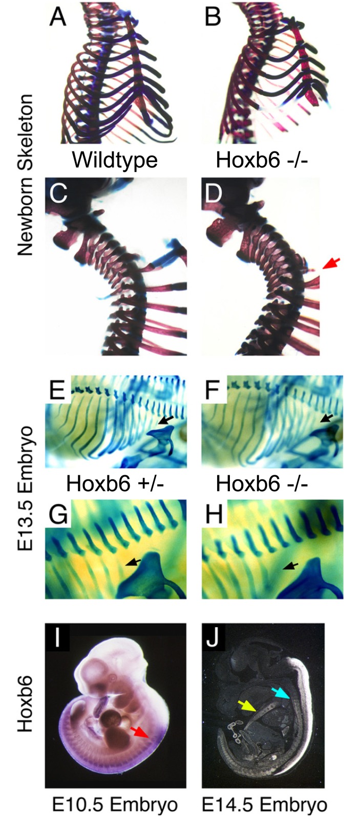
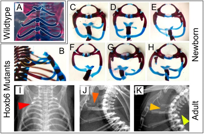
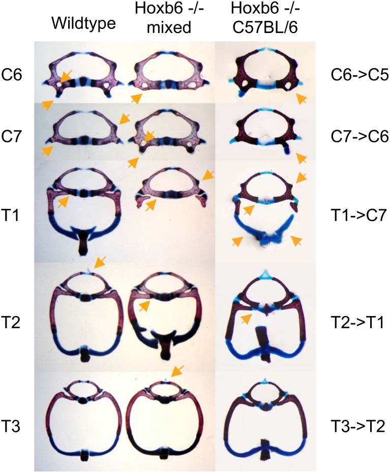
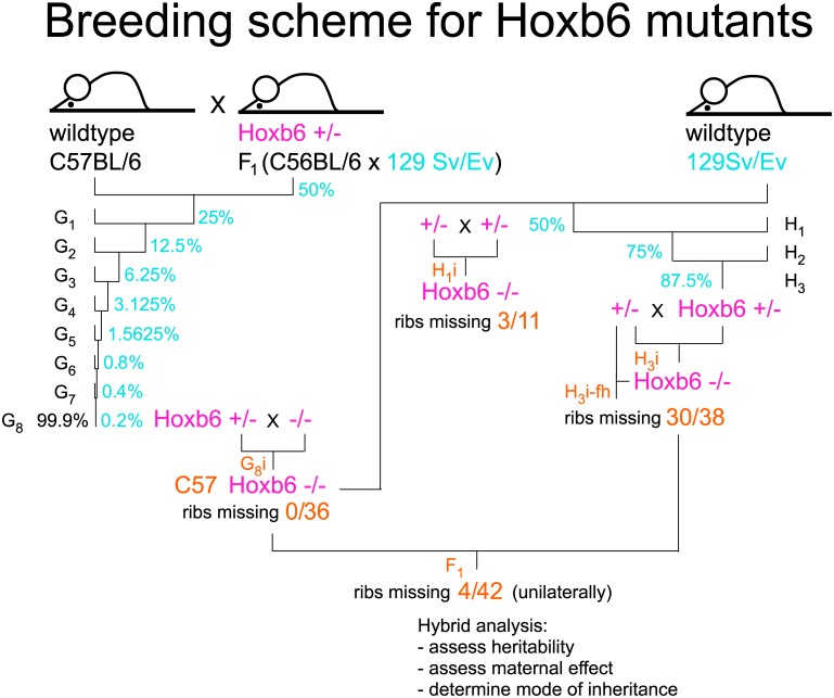
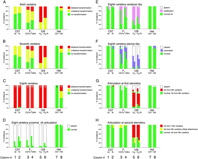
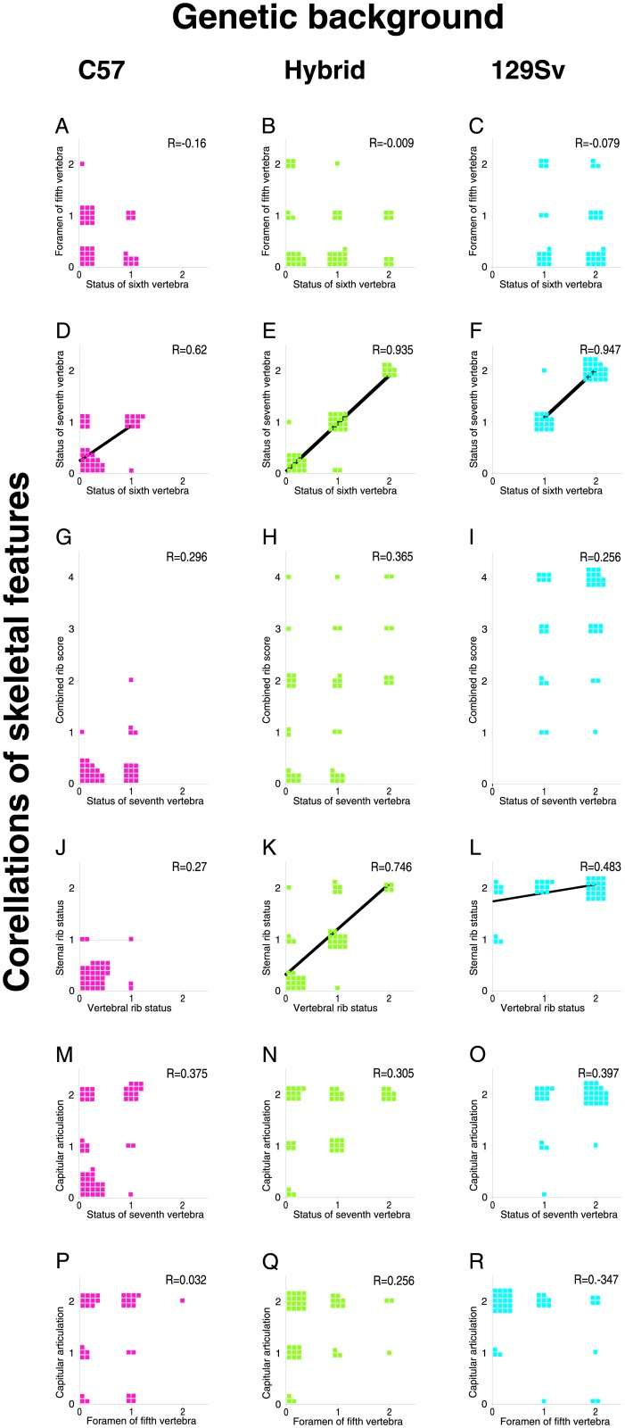
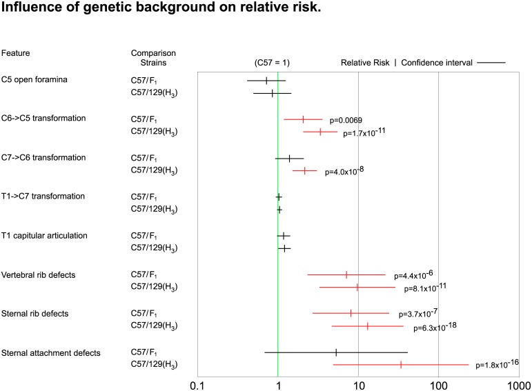
The process of patterning along the anterior-posterior axis in vertebrates is highly conserved. The function of Hox genes in the axis patterning process is particularly well documented for bone development in the vertebral column and the limbs. We here show that Hoxb6, in skeletal elements at the cervico-thoracic junction, controls multiple independent aspects of skeletal pattern, implicating discrete developmental pathways as substrates for this transcription factor. In addition, we demonstrate that Hoxb6 function is subject to modulation by genetic factors. These results establish Hox-controlled skeletal pattern as a quantitative trait modulated by gene-gene interactions, and provide evidence that distinct modifiers influence the function of conserved developmental genes in fundamental patterning processes.


Conflict of interest statement

Competing Interests: The author has declared that no competing interests exist.

Figures

Fig 1
Fig 1. Hoxb6 mutants exhibit skeletal alterations and absence of the first pair of ribs.
Panel A: Rib cage of wildtype skeleton with 7 ribs attached to the sternum and 6 sternebrae. Panel B: Rib cage of mutant with 6 ribs attached to sternum and 5 sternebrae. Panel C: Cervico-thoracic region of wildtype newborn skeleton. Panel D: Cervico-thoracic region of Hoxb6hd mutant newborn skeleton, the red arrow points to small ossified structures in place of the first pair of ribs. Panel E: E13.5 embryo heterozygous for the Hoxb6hd mutant allele stained with Alcian Blue reveals normal cartilage anlagen. Panel F: E13.5 homozygous Hoxb6hd mutant embryo with cartilage anlagen for the first rib absent (compare targets of black arrows). Panels G, H: Magnifications of Panels E and F, respectively. Panel I: Whole mount in situ hybridization for Hoxb6 in an embryo isolated at day E10.5. The anterior limit of Hoxb6 expression in somites is found within the caudal region of the somite that contributes to prevertebra 6 (red arrow). Panel J: In situ hybridization to a sagittal section from an embryo at E14.5 shows strong signal in spinal cord, and Hoxb6 expression is evident in the vertebral column (light blue arrow), and in the precursors to the sternum (yellow arrow).
Fig 2
Fig 2. Features of rib development and sternal articulation in Hoxb6hd mutants.
Panel A: Newborn wildtype skeleton with outlines (white) of cartilaginous portions of the ribs and sternum. Panels B-K: Hoxb6hd mutant skeletons from newborns (B-H) or adults (I-K). Panel B: Note the crossover of rib from the eighth vertebra and fusion with the rib from ninth vertebra, bifurcation of cartilage, and aberrant attachment of fused cartilage, and fusion of the first two sternebrae. Panels C-H: Different combinations in individuals of absent or rudimentary ribs, unilaterally (C-E) or bilaterally (F-H), defective formation of rib cartilage (D, E, G, H), crossovers (D, E, H), fusions (E, H), bifurcations (D, E, H), aberrant articulation to the sternum (C-H) and off-set sternal attachment of the ribs (H). All preparations contain the eighth and ninth vertebrae, except in Panel F, which represents the ninth vertebra. Panels I-K: X-rays of individual adult Hoxb6hd mutants. Panel I: frontal view; note truncation of first rib unilaterally (red arrow). Panel J: lateral view, orange arrow points to crossover and defective sternal rib cartilage. Panel K: lateral view, dark yellow arrow points to crossover and unilateral absence of first rib, bright yellow arrow points to absence of spinous process on the ninth vertebra.
Fig 3
Fig 3. Phenotype expressivity in Hoxb6 mutants on different genetic backgrounds.
Panels in column A: Vertebral elements of wildtype newborn mouse skeleton. Arrows point to characteristic features: Vertebra C6: vertebral foramen and anterior tuberculum; C7: lateral extension of the vertebral body and absence of foramen; T1: articulation of rib capitulum to vertebral body; T2: dorsal cartilage extension (processus spinosus). Panels in column B: Homeotic transformations in a homozygous Hoxb6hd mutant on mixed background. Arrows point to features found transposed to vertebrae at the next axial level, resembling anterior homeotic transformations of these skeletal elements. Panels in column C: Homeotic transformations in homozygous Hoxb6hd mutant on C57BL/6 background. Homeotic transformations are more often found unilaterally; arrow points to rib cartilage associated with the transformed eighth vertebra.
Fig 4
Fig 4. Breeding scheme for C57BL/6-Hoxb6hd congenic strain and backcross to 129S6/SvEv.
A male heterozygote for the Hoxb6hd mutation was crossed to C57BL/6 wildtype females and male offspring were used for further backcrosses. For the backcross to 129S6/SvEv wildtype, a Hoxb6hd homozygous male was used, and offspring from this cross (H1) were intercrossed (H1i) to generate mixed background animals homozygous for the Hoxb6 mutation. Out of 11 H1i progeny, 3 had defective first rib development, indicating an effect of the 129S6/SvEv genetic background on phenotype manifestation. Further backcrosses used H1 males heterozygous for the Hoxb6hd mutation and wildtype 129S6/SvEv females. Intercrosses of H3 animals (H3i) yielded homozygous Hoxb6hd mutants on predominantly 129S6/SvEv genetic background. To exclude a possible developmental disadvantage for the 25% homozygotes in a cross of Hoxb6hd heterozygous parents, we set up crosses between H3i generation animals in which the father was homozygous for the Hoxb6hd mutation (H3i-fh), thus increasing the yield of homozygous mutants to 50%, providing equal chance for intrauterine development. Of 38 mutant H3i progeny, 33 exhibited the “missing rib” phenotype, confirming the influence of genetic background on phenotype manifestation in Hoxb6hd mutants. Further backcrosses used homozygous mutant H1i progeny bred to C57BL/6-Hoxb6hd congenics (G8i), which produced 42 K2 progeny, of which 12 exhibited defective ribs; crosses of Hoxb6hd homozygous mutant F1 hybrids to H3 Hoxb6hd homozygous mutants produced 62 J2 animals, of which 55 had defective ribs. Differences in incidence of the phenotype are statistically significant between all groups (p = or < 0.01), except for the J2/H3 comparison. These results provide evidence that genetic background controls rib development in Hoxb6hd mutants in intermediate fashion.
Fig 5
Fig 5. Phenotype expressivity in Hoxb6 mutants is controlled by genetic background of the embryo.
Columns show the quantitative distribution of features by skeletal element and genetic background. Phenotype features were scored exactly as shown by orange arrows in Fig 3. Panel A: 6th vertebra; Panel B: 7th vertebra; Panel C: 8th vertebra (normally first thoracic vertebra); Panel D: Articulation of ribs/ossifications to the 8th vertebrae; Panel E: Proximal (vertebral) ribs; Panel F: Distal (sternal) ribs; Panel G: Articulation to sternum at level of 8th vertebra (normally T1); Panel H: Articulation to sternum at level of 9th vertebra (normally T2). For each experimental group, the fraction of animals with a given phenotype is plotted, and the total number of animals is given as a number in the respective columns of Panel C. No attempt was made to depict side of unilateral anomalies; as there was no preference, they were grouped together with increasing severity to the right of each column. Columns 1–8 correspond to the following groups: 1: Progeny from crosses of homozygous C57BL/6-Hoxb6hd mutant males to heterozygous C57BL/6-Hoxb6hd mutant females (G8i, see Fig 4) maintained on corn-cob bedding (C57 cb). 2: Progeny from crosses of homozygous C57BL/6-Hoxb6hd mutant males to heterozygous C57BL/6-Hoxb6hd mutant females (G8i) maintained on synthetic fiber bedding (C57 fb). 3: Progeny (F1) from crosses of homozygous H3 mutant males to homozygous C57BL/6-Hoxb6hd females (C57m: maternal uterine environment is C57BL/6). 4: Progeny (F1) from crosses of homozygous C57BL/6-Hox-6hd mutant males to homozygous H3 mutant females (129m: maternal uterine environment is predominantly 129Sv/Ev). 5: Progeny (H3i) from brother x sister matings of H3 generation animals on predominantly 129S6/SvEv background. 6: Progeny from crosses of homozygous H3 mutant males to heterozygous H3 mutant females (H3i-fh; homozygous father) on predominantly 129S6/SvEv background. 7: C57BL/6-Hoxb6hd heterozygous mutants (Het C57). 8: Heterozygous Hoxb6hd mutants from H3i intercrosses (Het 129). Statistical significance was established by 2-tailed Fisher’s exact test. For comparison of heterozygotes on the two different genetic backgrounds (Columns 7 and 8), p-values were not significant except for T1 status (p = 0.0095). For complete p-value matrices for all pairwise comparisons for columns 1–6 see Table 1.
Fig 6
Fig 6. Association of skeletal features in individual Hoxb6hd mutants.
Regression analyses were performed using a simplified scoring scheme for each character: two points were assigned for bilateral abnormalities, one point for unilateral abnormalities and 0 for wildtype manifestation. Each animal is represented by a square. The correlation coefficients (R) were determined using Microsoft Excel. Significance for relationships was assessed by ANOVA and was smaller than p = 0.05 (adjusted p<0.003 with correction for multiple comparisons) only for data in Panels D-F, K, L (solid regression curves). The presence of association indicates that the cellular and molecular processes producing a given feature are likely developmentally linked. The absence of relationship between skeletal anomalies within individual animals indicates that the various aspects of the Hoxb6hd phenotype develop independently from each other, possibly controlled by different underlying molecular mechanisms.
Fig 7
Fig 7. Influence of genetic background on risk for skeletal abnormalities in Hoxb6 mutants.
The fraction of animals with abnormalities (uni- and bilateral combined) for each group was determined and risk was calculated relative to C57BL/6-Hoxb6hd homozygous mutants. Relative Risk increases with increasing contribution from the 129S6/SvEv genetic background; for some features, this increase is significant (p<0.05) in both chi-square and Fisher's exact test (p-values are shown). Confidence intervals (horizontal bars) were determined for the comparisons of relative risk estimate (small vertical bar) for Hoxb6 mutants on F1 hybrid and 129S6/SvEv genetic background, respectively, based upon the results in S2 Table. Relative risk for anomalies in Hoxb6hd mutants on C57BL/6 background was set to 1 (green line), and risk estimates whose confidence intervals do NOT include the value 1 are highly significant as indicated by red color of the data points.

Similar articles

Cited by

References

    1. Capecchi MR. Hox genes and mammalian development. Cold Spring Harb Symp Quant Biol. 1997;62:273–281. - PubMed
    1. Davis AP, Capecchi MR. Axial homeosis and appendicular skeleton defects in mice with a targeted disruption of hoxd-11. Development. 1994;120:2187–2198. - PubMed
    1. Kostic D, Capecchi MR. Targeted disruptions of the murine hoxa-4 and hoxa-6 genes result in homeotic transformations of components of the vertebral column. Mechanisms of Development. 1994;46:231–247. - PubMed
    1. Ramirez-Solis R, Zheng H, Whiting J, Krumlauf R, Bradley A. Hoxb-4 (hox-2.6) mutant mice show homeotic transformation of a cervical vertebra and defects in the closure of the sternal rudiments. Cell. 1993;73:279–294. - PubMed
    1. Rancourt DE, Tsuzuki T, Capecchi MR. Genetic interaction between hoxb-5 and hoxb-6 is revealed by nonallelic noncomplementation. Genes Dev. 1995;9:108–122. - PubMed

Publication types