. 2016 Jan 22;11(1):e0147236.
doi: 10.1371/journal.pone.0147236. eCollection 2016.

Expression of the Long Non-Coding RNA HOTAIR Correlates with Disease Progression in Bladder Cancer and Is Contained in Bladder Cancer Patient Urinary Exosomes

Affiliations

Expression of the Long Non-Coding RNA HOTAIR Correlates with Disease Progression in Bladder Cancer and Is Contained in Bladder Cancer Patient Urinary Exosomes

Claudia Berrondo et al. PLoS One. .

Abstract

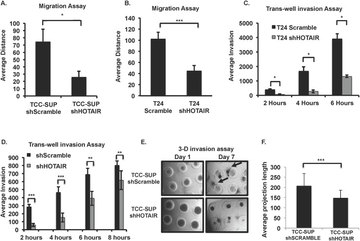
Exosomes are 30-150nM membrane-bound secreted vesicles that are readily isolated from biological fluids such as urine (UEs). Exosomes contain proteins, micro RNA (miRNA), messenger RNA (mRNA), and long non-coding RNA (lncRNA) from their cells of origin. Although miRNA, protein and lncRNA have been isolated from serum as potential biomarkers for benign and malignant disease, it is unknown if lncRNAs in UEs from urothelial bladder cancer (UBC) patients can serve as biomarkers. lncRNAs are > 200 nucleotide long transcripts that do not encode protein and play critical roles in tumor biology. As the number of recognized tumor-associated lncRNAs continues to increase, there is a parallel need to include lncRNAs into biomarker discovery and therapeutic target algorithms. The lncRNA HOX transcript antisense RNA (HOTAIR) has been shown to facilitate tumor initiation and progression and is associated with poor prognosis in several cancers. The importance of HOTAIR in cancer biology has sparked interest in using HOTAIR as a biomarker and potential therapeutic target. Here we show HOTAIR and several tumor-associated lncRNAs are enriched in UEs from UBC patients with high-grade muscle-invasive disease (HGMI pT2-pT4). Knockdown of HOTAIR in UBC cell lines reduces in vitro migration and invasion. Importantly, loss of HOTAIR expression in UBC cell lines alters expression of epithelial-to-mesenchyme transition (EMT) genes including SNAI1, TWIST1, ZEB1, ZO1, MMP1 LAMB3, and LAMC2. Finally, we used RNA-sequencing to identify four additional lncRNAs enriched in UBC patient UEs. These data, suggest that UE-derived lncRNA may potentially serve as biomarkers and therapeutic targets.


Conflict of interest statement

Competing Interests: The authors have declared that no competing interests exist.

Figures

Fig 1
Fig 1. UBC cell lines over-express tumor-associated lncRNA and mRNA relative to the SV-HUC non-tumorigenic urothelial cell line.
qRT-PCR was performed on cell lysates from a range of UBC cell lines and the control SV-40 immortalized urothelial cell line SV-HUC. The level of mRNA and lncRNA expression was first normalized to GAPDH and fold-change of gene expression in individual UBC cell lines was compared to the control SV-HUC cells. (A) T24 cell lysates (B) TCC-SUP cell lysates. (C) qRT-PCR of HOTAIR in UBC cell lines normalized to SV-HUC cell line transcript levels. (n = 3 experiments). Student’s t-tests were used for all experiments to identify statistical significance in expression between UBC and control SV-HUC cells (Fig 1A–1C) * p<0.05, ** p<0.001, ***p<0.0001.
Fig 2
Fig 2. HOTAIR knockdown affects in vitro migration and invasion in UBC cells.
Distance of migration was measured following scratch-wound assay of lentiviral shRNA knockdown of HOTAIR vs. control shScramble (A) T24 and (B) TCC-SUP UBC cell lines. Trans-well invasion assay of shHOTAIR vs. control shScramble (C) T24 and (D). TCC-SUP cell lines (E) 3-D invasion assay comparing shScramble control TCC-SUP cells to shHOTAIR TCC-SUP cells. Cells are seeded into microtissue ® generated caster gels and allowed to form spheroids. After spheroids are formed, BME is gently layered over the caster gel. Dark circular spheroids are shown and arrows point to projections of invading cells into the surrounding BME. F. Following 7 days of culture, projection lengths were measured from spheroid surface to the distal tip using ImageJ and the average length of projection determined for each cell type. Student’s t-test was used to determine statistical differences in each experiment presented (A-F) *p<0.1, **p<0.05, ***p<0.01 (n = 3–6 experiments/panel).
Fig 3
Fig 3. Loss of HOTAIR affects EMT factor expression in UBC cell lines.
(A) qRT-PCR of known HOTAIR target genes and classical EMT factors mRNA in shHOTAIR T24 compared to shScramble T24 UBC cells. (B) EMT target gene mRNA expression in shHOTAIR TCC-SUP cells compared to shScramble TCC-SUP cells (in panels A and B EMT mRNA levels were normalized to GAPDH). (C) siRNA targeted against HOTAIR or GFP was used in T24 cells and EMT factors ZEB1 and SNAI1 mRNA expression evaluated by qRT-PCR (mRNA was normalized to 18s). (D) Immunoblot of T24 siGFP and siHOTAIR cells showing reduced protein levels of ZEB1 and SNAI1. GAPDH is a loading control. Beta-actin was used as a loading control. Student’s t-test was used to determine statistical differences between control and HOTAIR knockdown cells in the qRT-PCR experiments presented (A-C) *p<0.1, **p<0.05, ***p<0.01 (n = 3–6 experiments/panel).
Fig 4
Fig 4. HGMI (pT2-pT4) UBC patient tumors overexpress several tumor-associated lncRNA and mRNA.
(A) qRT-PCR of tumor-associated mRNAs and lncRNAs in tumors from patients who underwent cystectomy for HG disease (final pathology pT2-pT4). Tumor and distal normal tissue (DNT) mRNA or lncRNA was normalized to 18s and then the ratio of tumor to DNT was calculated. Statistical significance was determined using Student’s t-test to compare tumor to DNT normalized expression, *p<0.05. (n = 10 patients). (B) HOTAIR was not differentially expressed between pT2 tumors and pT4 tumors (Student’s t-test, p>0.05).
Fig 5
Fig 5. Tumor-associated lncRNAs and mRNAs are enriched in T24 and TCC-SUP exosomes relative to control cell line exosomes.
(A) Electron micrograph of exosomes isolated from T24 and TCC-SUP UBC cell lines. White bar 100nm. (B) Particle analysis of TCC-SUP exosomes using the LM10 nanoparticle characterization system (NanoSight). Particles were observed in the size range of 30-150nm (n = 3 experiments). (C) Western blot of exosomes isolated from TCC-SUP UBC cell line. ALIX is a well-characterized exosome marker and GAPDH is a loading control. (D-E) The level of exosomal mRNA and lncRNA expression was normalized to 18S and fold-change of gene expression in T24 and TCC-SUP exosomes was compared to control SV-HUC cell exosomes (D) T24 exosomes and (E) TCC-SUP exosomes. (n = 3 for each experiment). Student’s T-test was used to determine statistical significance between UBC and control exosomes (*p<0.1, **p<0.05, ***p<0.01). (F-G) UBC cell line exosomes are enriched in tumor-associated lncRNAs and mRNAs relative to their producer cell lysates. The ratio of 18S-normalized exosome to cell lysate lncRNAs and mRNAs is shown for (F) T24 and (G) TCC-SUP.
Fig 6
Fig 6. Exosomes isolated from the urine of HGMI (pT2-pT4) UBC patients are enriched in tumor-associated lncRNA and mRNA.
(A) Western blot of UEs from five patients. ALIX is a well-known exosome marker, and GAPDH is a loading control. (B) Representative electron micrograph of exosomes purified from the urine of a UBC patient with HGMI disease (pT2-pT4 final pathology). (C) qRT-PCR of UEs for tumor-associated mRNA and lncRNA from UBC patients (n = 8) and HVs (n = 5) and converted to cDNA. qRT-PCR was performs for mRNA and lncRNA. Patient samples were normalized to 18s and HVs normalized to 18s. Student’s t-test **p<0.01 and ***p < .001.
Fig 7
Fig 7. lncRNAs HYMA1, LINC00477, LOC100506688 and OTX2-AS1 are enriched in UEs of UBC patients with HGMI disease (pT2-pT4).
Confirmatory qRT-PCR of the novel lncRNAs (normalized to 18s) identified by RNA-sequencing in n = 8 UEs from patients with HGMI (pT2-pT4) UBC and UEs from n = 7 HVs. Student’s t-test *p<0.05, **p<0.01, ***p<0.001.

References

    1. Fatica A, Bozzoni I. Long non-coding RNAs: new players in cell differentiation and development. Nat Rev Genet. 2014;15: 7–21. 10.1038/nrg3606 - DOI - PubMed
    1. Fatima R, Akhade VS, Pal D, Rao SM. Long noncoding RNAs in development and cancer: potential biomarkers and therapeutic targets. Mol Cell Ther. 2015;3: 5 10.1186/s40591-015-0042-6 - DOI - PMC - PubMed
    1. Gupta RA, Shah N, Wang KC, Kim J, Horlings HM, Wong DJ, et al. Long non-coding RNA HOTAIR reprograms chromatin state to promote cancer metastasis. Nature. 2010;464: 1071–1076. 10.1038/nature08975 - DOI - PMC - PubMed
    1. Doose G, Haake A, Bernhart SH, López C, Duggimpudi S, Wojciech F, et al. MINCR is a MYC-induced lncRNA able to modulate MYC’s transcriptional network in Burkitt lymphoma cells. Proc Natl Acad Sci U S A. 2015;112: E5261–70. - PMC - PubMed
    1. Sun M, Nie F-Q, Wang Z-X, De W. Involvement of lncRNA dysregulation in gastric cancer. Histol Histopathol. 2016;31: 33–9. 10.14670/HH-11-655 - DOI - PubMed

Publication types