. 2016 May;27(5):400-15.
doi: 10.1089/hum.2015.126. Epub 2016 Mar 30.

Conditional Cytotoxic Anti-HIV Gene Therapy for Selectable Cell Modification

Affiliations

Conditional Cytotoxic Anti-HIV Gene Therapy for Selectable Cell Modification

Himanshu Garg et al. Hum Gene Ther. 2016 May.

Abstract

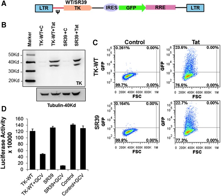
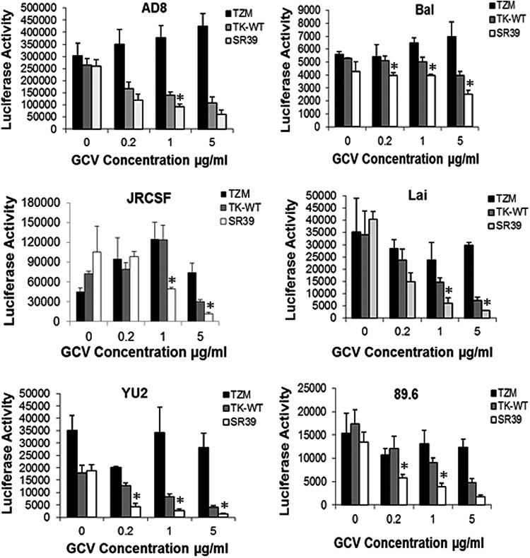
Gene therapy remains one of the potential strategies to achieve a cure for HIV infection. One of the major limitations of anti-HIV gene therapy concerns recovering an adequate number of modified cells to generate an HIV-proof immune system. Our study addresses this issue by developing a methodology that can mark conditional vector-transformed cells for selection and subsequently target HIV-infected cells for elimination by treatment with ganciclovir (GCV). We used the herpes simplex virus thymidine kinase (TK) mutant SR39, which is highly potent at killing cells at low GCV concentrations. This gene was cloned into a conditional HIV vector, pNL-GFPRRESA, which expresses the gene of interest as well as green fluorescent protein (GFP) in the presence of HIV Tat protein. We show here that TK-SR39 was more potent that wild-type TK (TK-WT) at eliminating infected cells at lower concentrations of GCV. As the vector expresses GFP in the presence of Tat, transient expression of Tat either by Tat RNA transfection or transduction by a nonintegrating lentiviral (NIL) vector marked the cells with GFP for selection. In cells selected by this strategy, TK-SR39 was more potent at limiting virus replication than TK-WT. Finally, in Jurkat cells modified and selected by this approach, infection with CXCR4-tropic Lai virus could be suppressed by treatment with GCV. GCV treatment limited the number of HIV-infected cells, virus production, as well as virus-induced cytopathic effects in this model. We provide proof of principle that TK-SR39 in a conditional HIV vector can provide a safe and effective anti-HIV strategy.


Figures

<b>Figure 1.</b>
Figure 1.
The thymidine kinase mutant SR39 (TK-SR39) construct shows significantly higher cell killing compared with its wild-type counterpart (TK-WT). (A) HeLa cells were transfected with the TK-WT or TK-SR39 gene cloned into the mammalian pcDNA3.1 expression vector or with empty pcDNA3.1 vector as control. Cells were then cultured in the presence of various concentrations of ganciclovir (GCV) as indicated. Cell viability was determined 72 hr posttransfection, using the CellTiter-Glo viability assay (Promega). Data represent means ± SD of triplicate observations. One representative of three independent experiments is shown. (B) Both vectors expressed the TK gene to similar levels as evident by Western blot analyses using antibodies against the TK protein.
<b>Figure 2.</b>
Figure 2.
TK-WT and TK-SR39 genes cloned in the conditional vector pGFPRRESA express the TK gene and green fluorescent protein (GFP) in the presence of HIV Tat and inhibit HIV production in the presence of GCV. (A) Schematic of Tat-dependent lentiviral vector used in the study. HeLa cells were transfected with the TK-WT or TK-SR39 gene expressed by the conditional vector pGFPRRESA in the presence or absence of viral protein Tat, using the Tat expression vector. (B) Expression of the TK gene was determined 48 hr posttransfection by Western blotting, using antibodies against the TK protein. (C) GFP expression in the presence or absence of HIV Tat was determined by flow cytometric analysis. (D) HeLa cells were cotransfected with HIV pNL4-3 along with TK-WT, TK-SR39, or empty lentiviral control vector. GCV at a concentration of 10 μg/ml was added to the respective wells, and culture supernatants were harvested and used to infect TZM cells. Luciferase activity was determined in infected TZM cells 24 hr postinfection, using the britelite luciferase assay substrate as a direct measure of infection. Columns represent means and SD of triplicate observations. C, control; LTR, long terminal repeat; IRES, internal ribosomal entry site; RRE, Rev response element. Color images available online at www.liebertpub.com/hum
<b>Figure 3.</b>
Figure 3.
Tat RNA can be used to sort pGFPRRESA-TK-WT or pGFPRRESA-TK-SR39 lentiviral vector-transduced cells. (A) TZM-bl cells were transfected with various concentrations of Tat DNA or RNA followed by determination of luciferase activity 24 hr posttransfection. Columns represent means and SD of triplicate observations. (B) TZM-bl cells were infected with TK-WT- or TK-SR39-packaged virion particles followed by transfection with Tat RNA. The cells were then assayed for GFP expression followed by sorting of GFP-positive cells. Color images available online at www.liebertpub.com/hum
<b>Figure 4.</b>
Figure 4.
Sorted TZM-bl cells maintain dormant integrated lentiviral vector in a stable form. GFP-positive cells transduced with TK-WT or TK-SR39 lentiviral vector and sorted as described in Fig. 3 were expanded in cell culture for approximately 3–4 weeks. (A) Cells were retransfected with Tat RNA or transduced with NL-Luc followed by determination of GFP expression by flow cytometry. (B) Expression of the TK gene in control (C), Tat-transfected, or NL-Luc-infected cells was determined by Western blotting with an anti-TK antibody. (C) The above-sorted cells were maintained in culture for approximately 8 months (240 days) and then infected with NL-Luc. TK-SR39 cells maintained the TK gene in a stable form as evident by GFP expression.
<b>Figure 5.</b>
Figure 5.
Lentivirally expressed TK-SR39 construct shows significantly higher cell killing in the presence of lower GCV concentrations. Sorted TZM cells transduced with the TK-WT or TK-SR39 vector were either transfected with (A) pcDNA or (B) Tat DNA, or (C) infected with pNL-Luc/VSVG. GCV was added at the respective concentrations and cell viability was determined 48 hr posttreatment, using the CellTiter-Glo viability assay. Viability was normalized to 100% in the absence of GCV. Data represent means ± SD of triplicate observations. One representative of three independent experiments is shown.
<b>Figure 6.</b>
Figure 6.
HIV inhibition in cells lentivirally transduced with TK-WT or TK-SR39. Parental TZM cells or sorted TZM cells transduced with the TK-WT or TK-SR39 vector were infected with various HIV isolates. Four to 5 hr postinfection, culture medium was replaced with RPMI containing the indicated concentrations of GCV. Culture supernatants were harvested on day 3 (AD8, Bal, Lai, YU-2, 89.6) or day 6 (JRCSF) postinfection and used to infect TZM cells that were analyzed for luciferase activity 24 hr postinfection. Data represent means and SD of triplicate observations. *Significant difference (p < 0.05) in HIV production in TK-SR39-transduced cells compared with TK-WT.
<b>Figure 7.</b>
Figure 7.
The Tat-NIL (nonintegrating lentiviral) vector can be used to effectively sort TK-WT- and TK-SR39-transduced cells. (A) Sorted TZM-TK-SR39 cells were infected with Tat-NIL-packaged lentiviral particles. The cells were monitored for GFP expression over a period of 15 days. GFP packaged by an integrating lentiviral vector was used as control. (B) Jurkat cells were infected with TK-WT or TK-SR39 lentiviral particles followed by infection with Tat-NIL lentiviral particles. Cells were then analyzed for GFP expression on day 3 postinfection followed by sorting of GFP+ cells. Jurkat cells sorted in (B) were expanded in culture, infected with NL-Luc, and analyzed for (C) GFP expression by flow cytometry. (D) Expression of the TK gene as determined by Western blotting. (E) Sorted Jurkat T cells transduced with the TK-WT or TK-SR39 vector were either left uninfected or infected with pNL-Luc/VSVG. GCV was added at the respective concentrations and cell viability was determined 48 hr posttreatment, using the CellTiter-Glo viability assay. (F) CD34+ stem cells were transduced with TK-SR39 vector followed by transduction with Tat-NIL the next day. Cells were analyzed for GFP expression 48 hr later by flow cytometry and fluorescence microscopy analysis.
<b>Figure 8.</b>
Figure 8.
TK-SR39-transduced cultures specifically deplete HIV-infected cells in the presence of GCV. Sorted TK-WT- or TK-SR39-transduced Jurkat cells were infected with VSV pseudotyped NL-LUC followed by culture in the presence of various concentrations of GCV. On days 5 and 7, cells were stained with propidium iodide (PI) and analyzed by flow cytometry. (A) Contour plots showing the percentage of HIV-infected cells undergoing cell death (GFP+PI+) or not (GFP+PI) in the presence or absence of GCV. (B) Analysis of GFP+PI+ or GFP+PI cells among Jurkat-TK-WT or Jurkat-SR39 cells in the presence of various GCV concentrations. The horizontal line shows the levels of HIV-infected but GCV-untreated cells.
<b>Figure 9.</b>
Figure 9.
TK-SR39-modified cells inhibit HIV replication in the presence of GCV, allowing for preservation of cell viability in infected cultures. Parental Jurkat cells or Jurkat cells lentivirally transduced with TK-WT or TK-SR39 and then sorted were infected with HIV-Lai virus followed by culture in the presence of the indicated concentrations of GCV. (A) Culture supernatants were harvested on the indicated days postinfection and used to infect TZM cells as a direct measure of infectious virus in cultures after respective treatments. (B) Culture supernatants were harvested on the indicated days postinfection to determine reverse transcriptase (RT) activity as a measure of virus replication. (C) Cell viability was determined on day 5, 7, or 10 postinfection, using the CellTiter-Glo viability assay. Percent viability was calculated after normalizing to uninfected controls. (D) A direct comparison of inhibition of infectious HIV production in parental Jurkat, Jurkat-TK-WT, or Jurkat-SR39 cells over a period of 17 days after treatment with GCV at a concentration of 20 μg/ml, using data from (A). (E) Infected cells were visualized by microscopy for virus-induced cytopathic effects (syncytium formation) and GFP expression.
<b>Figure 10.</b>
Figure 10.
TK-SR39-modified cells inhibit HIV replication by killing HIV-infected cells accompanied by preservation of the live cell population. Sorted TK-WT- or TK-SR39-transduced Jurkat cells were infected with wild-type HIV-Lai virus followed by culture in the presence of various GCV concentrations. (A) GFP-positive cells as an indicator of HIV infection were monitored by flow cytometry over a period of 10 days. (B) Population statistics of the live-versus-dead cell populations in uninfected or HIV-infected TK-WT- or TK-SR39-transduced cells in the presence (20 μg/ml) or absence of GCV. (C) Flow cytometry-based analysis of the percentage of live versus dead cell populations in sorted TK-SR39-transduced Jurkat cells in the presence of the indicated GCV concentrations. Color images available online at www.liebertpub.com/hum
<b>Figure 11.</b>
Figure 11.
Overall schematic of strategy for generation of genetically modified stem cells, using the TK-SR39 lentiviral vector, and the outcome of genetically modified cells on HIV infection. The conditional cytotoxic lentiviral vector pNL-GFPRRESA-TK-SRR9 expresses the TK gene only in the presence of HIV Tat. On Tat expression, the cells also express GFP. The strategy can be exploited in vitro for sorting/enrichment of genetically modified cells. During HIV infection, Tat is provided by the virus leading to TK-SR39 gene expression. When treated with GCV, the TK-expressing, HIV-infected cells are marked for elimination by apoptosis. Color images available online at www.liebertpub.com/hum

References

    1. Hutter G, and Ganepola S. Eradication of HIV by transplantation of CCR5-deficient hematopoietic stem cells. ScientificWorldJournal 2011;11:1068–1076 - PMC - PubMed
    1. Hutter G, Nowak D, Mossner M, et al. . Long-term control of HIV by CCR5 Δ32/Δ32 stem-cell transplantation. N Engl J Med 2009;360:692–698 - PubMed
    1. Younan P, Kowalski J, and Kiem HP. Genetic modification of hematopoietic stem cells as a therapy for HIV/AIDS. Viruses 2013;5:2946–2962 - PMC - PubMed
    1. Younan P, Kowalski J, and Kiem HP. Genetically modified hematopoietic stem cell transplantation for HIV-1-infected patients: can we achieve a cure? Mol Ther 2014;22:257–264 - PMC - PubMed
    1. Bennett MS, and Akkina R. Gene therapy strategies for HIV/AIDS: preclinical modeling in humanized mice. Viruses 2013;5:3119–3141 - PMC - PubMed

Publication types

MeSH terms

Substances