Coexistence of congenital diaphragmatic hernia and abdominal wall closure defect with chromosomal abnormality: two case reports
- PMID: 26800685
- PMCID: PMC4724109
- DOI: 10.1186/s13256-016-0805-y
Coexistence of congenital diaphragmatic hernia and abdominal wall closure defect with chromosomal abnormality: two case reports
Abstract
Background: We reported two rare cases of congenital diaphragmatic hernia with abdominal wall closure defect, which were not associated with septum transversum diaphragmatic defects or Fryns syndrome.
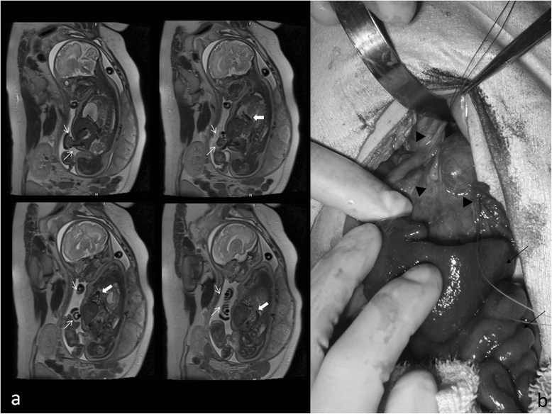
Case presentation: Case 1: a Japanese baby boy was delivered at 37 weeks' gestation by urgent cesarean section because of the diagnosis of severe fetal distress. Congenital diaphragmatic hernia with omphalocele was prenatally diagnosed with fetal ultrasound. A ruptured omphalocele was confirmed at delivery. A silo was established on the day of his birth; direct closure of his diaphragmatic defect and abdominal wall closure was performed on the fifth day after his birth. Trisomy 13 was confirmed by genetic examination. His postoperative course was uneventful and he was discharged 5 months postnatally with home oxygen therapy. He was readmitted because of heart failure and died at 6 months. Case 2: a Japanese baby boy, who was prenatally diagnosed with gastroschisis, was delivered at 35 weeks' gestation by urgent cesarean section because of the diagnosis of fetal distress. Silo construction using a wound retractor was performed on the day of his birth and direct abdominal closure was performed on the tenth day after his birth. Trisomy 21 was confirmed by genetic examination. Treatment for his respiratory distress was continued after surgery. A retrosternal hernia was revealed at 6 months and direct closure of retrosternal diaphragm with the resection of hernia sac was performed. His postoperative course was uneventful and he was discharged with home oxygen therapy.
Conclusions: Attention should be paid to chromosomal abnormality in cases in which the coexistence of congenital diaphragmatic hernia and abdominal wall closure defect are observed.
Figures


Similar articles
-
Abdominal wall defects and congenital heart disease.Ultrasound Obstet Gynecol. 2003 Apr;21(4):334-7. doi: 10.1002/uog.93. Ultrasound Obstet Gynecol. 2003. PMID: 12704739
-
Prenatal imaging of a fetus with the rare combination of a right congenital diaphragmatic hernia and a giant omphalocele.Congenit Anom (Kyoto). 2014 Nov;54(4):246-9. doi: 10.1111/cga.12075. Congenit Anom (Kyoto). 2014. PMID: 25059273
-
Prenatal and postnatal management of a patient with pentalogy of Cantrell and left ventricular aneurysm. A case report and literature review.Fetal Diagn Ther. 2007;22(4):269-73. doi: 10.1159/000100788. Epub 2007 Mar 16. Fetal Diagn Ther. 2007. PMID: 17369693 Review.
-
Prenatal diagnosis of Cantrell's pentalogy with conventional and three-dimensional sonography.J Matern Fetal Neonatal Med. 2002 Sep;12(3):209-11. doi: 10.1080/jmf.12.3.209.211. J Matern Fetal Neonatal Med. 2002. PMID: 12530621
-
Prenatal diagnosis and array comparative genomic hybridization characterization of trisomy 21 in a fetus associated with right congenital diaphragmatic hernia and a review of the literature of chromosomal abnormalities associated with congenital diaphragmatic hernia.Taiwan J Obstet Gynecol. 2015 Feb;54(1):66-70. doi: 10.1016/j.tjog.2014.12.001. Taiwan J Obstet Gynecol. 2015. PMID: 25675923 Review.
Cited by
-
A clinical-pathogenetic approach on associated anomalies and chromosomal defects supports novel candidate critical regions and genes for gastroschisis.Pediatr Surg Int. 2018 Sep;34(9):931-943. doi: 10.1007/s00383-018-4331-4. Epub 2018 Aug 9. Pediatr Surg Int. 2018. PMID: 30094464 Review.
-
Omphalocele and Associated Anomalies: Exploring Pulmonary Development and Genetic Correlations-A Literature Review.Diagnostics (Basel). 2025 Mar 10;15(6):675. doi: 10.3390/diagnostics15060675. Diagnostics (Basel). 2025. PMID: 40150018 Free PMC article. Review.
-
Left congenital diaphragmatic hernia and gastroschisis in a term male infant.BMJ Case Rep. 2021 Jul 22;14(7):e239181. doi: 10.1136/bcr-2020-239181. BMJ Case Rep. 2021. PMID: 34301696 Free PMC article.
-
Rare combination of left-sided congenital diaphragmatic hernia and omphalocele.BMJ Case Rep. 2017 Aug 7;2017:bcr2017220696. doi: 10.1136/bcr-2017-220696. BMJ Case Rep. 2017. PMID: 28790097 Free PMC article.
References
-
- Nonaka A, Hidaka N, Kido S, Fukushima K, Kato K. Prenatal imaging of a fetus with the rare combination of a right congenital diaphragmatic hernia and a giant omphalocele. Congenit Anom. 2014;54:246–9. - PubMed
Publication types
MeSH terms
LinkOut - more resources
Full Text Sources
Other Literature Sources
Medical

