. 2016 Mar;202(3):1167-83.
doi: 10.1534/genetics.115.185371. Epub 2016 Jan 22.

Hedgehog Signaling Strength Is Orchestrated by the mir-310 Cluster of MicroRNAs in Response to Diet

Affiliations

Hedgehog Signaling Strength Is Orchestrated by the mir-310 Cluster of MicroRNAs in Response to Diet

Ibrahim Ömer Çiçek et al. Genetics. 2016 Mar.

Abstract

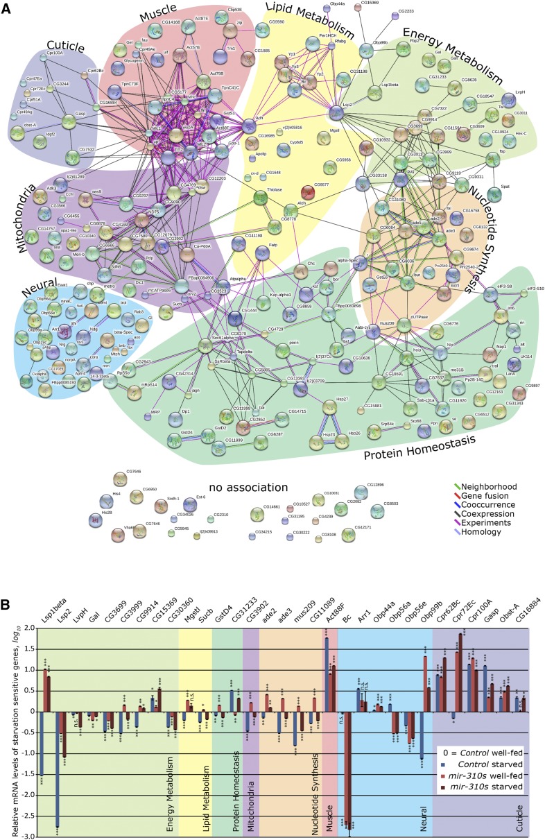
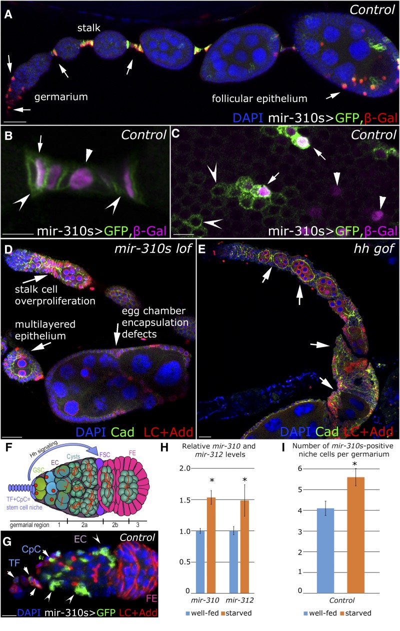
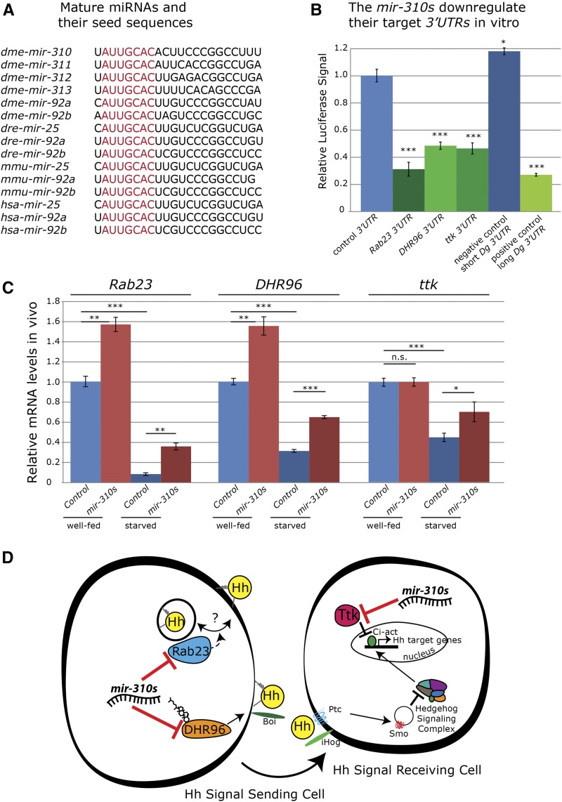
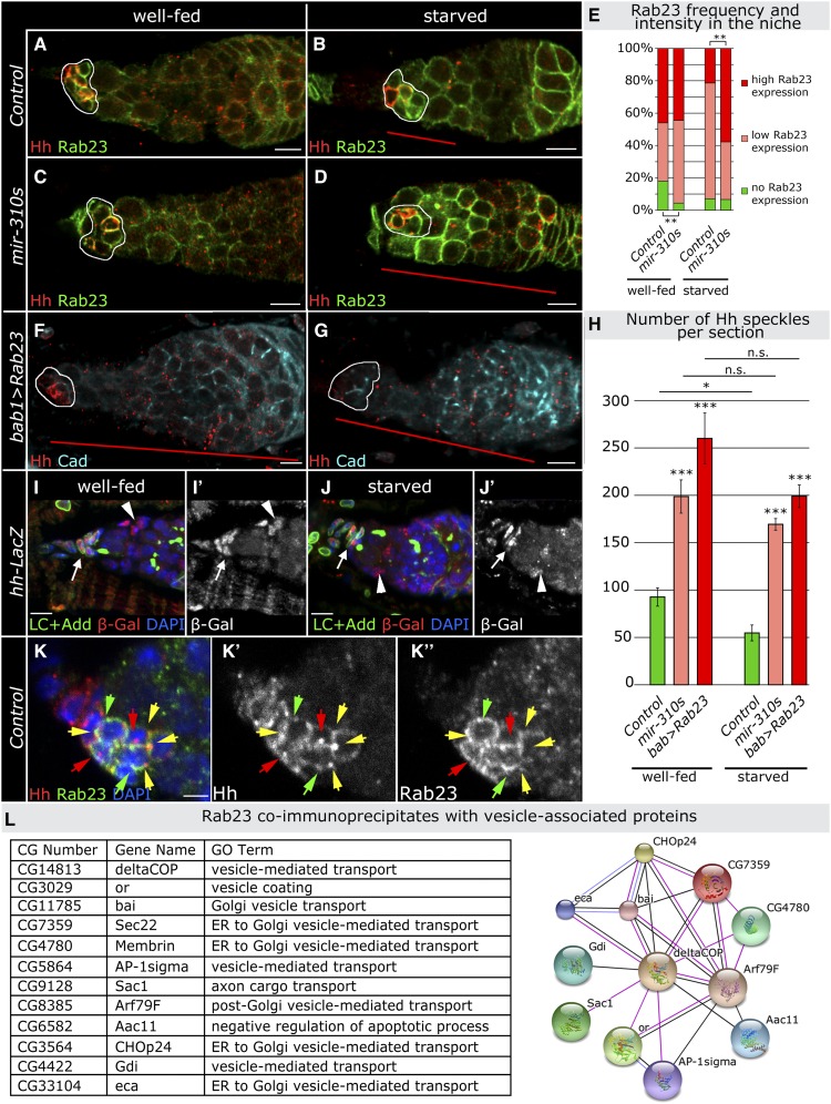
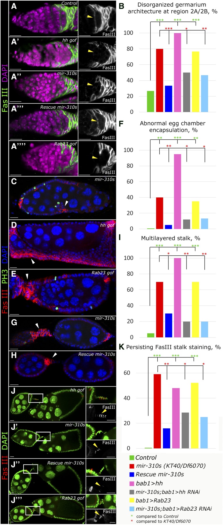
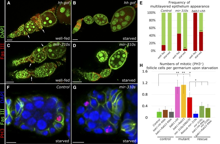
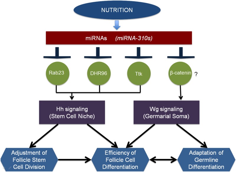
Since the discovery of microRNAs (miRNAs) only two decades ago, they have emerged as an essential component of the gene regulatory machinery. miRNAs have seemingly paradoxical features: a single miRNA is able to simultaneously target hundreds of genes, while its presence is mostly dispensable for animal viability under normal conditions. It is known that miRNAs act as stress response factors; however, it remains challenging to determine their relevant targets and the conditions under which they function. To address this challenge, we propose a new workflow for miRNA function analysis, by which we found that the evolutionarily young miRNA family, the mir-310s (mir-310/mir-311/mir-312/mir-313), are important regulators of Drosophila metabolic status. mir-310s-deficient animals have an abnormal diet-dependent expression profile for numerous diet-sensitive components, accumulate fats, and show various physiological defects. We found that the mir-310s simultaneously repress the production of several regulatory factors (Rab23, DHR96, and Ttk) of the evolutionarily conserved Hedgehog (Hh) pathway to sharpen dietary response. As the mir-310s expression is highly dynamic and nutrition sensitive, this signal relay model helps to explain the molecular mechanism governing quick and robust Hh signaling responses to nutritional changes. Additionally, we discovered a new component of the Hh signaling pathway in Drosophila, Rab23, which cell autonomously regulates Hh ligand trafficking in the germline stem cell niche. How organisms adjust to dietary fluctuations to sustain healthy homeostasis is an intriguing research topic. These data are the first to report that miRNAs can act as executives that transduce nutritional signals to an essential signaling pathway. This suggests miRNAs as plausible therapeutic agents that can be used in combination with low calorie and cholesterol diets to manage quick and precise tissue-specific responses to nutritional changes.

Keywords: Drosophila; Hedgehog signaling; Hh ligand; Rab23; dietary restriction; follicle stem cell; metabolic stress; miRNA; oogenesis; the mir-310s.


Figures

Figure 1
Figure 1
The mir-310s-affected genes suggest energy metabolism-related defects. (A) Interaction network of globally up- or downregulated genes in mir-310s loss-of-function (KT40/KT40) mutant females compared to control (w1118) females exhibits eight interconnected gene ontology groups: energy metabolism, lipid metabolism, protein homeostasis, muscle and neural development and function, mitochondrial, nucleotide synthesis, and cuticle related (Table S1). Large node size indicates the availability of protein structure information. Node colors have no particular meaning. Line colors indicate different evidence types used to generate node interactions (see key). (B) Starvation-sensitive genes have altered mRNA expression levels in mir-310s mutant females compared to controls under well-fed and/or nutritionally restricted conditions (10 days), demonstrating the role of the mir-310s in the response to changes in nutritional status (Table S2). Functional groups are color coded as in A. In B the bar graph indicates the arithmetic mean (AVE) ± the standard error of the mean (SEM). Significances were calculated using two-tailed Student’s t-test. *P < 0.05, **P < 0.005, ***P < 0.0005.
Figure 2
Figure 2
The mir-310s are expressed in the ovarian soma. (A–C) The mir-310s are expressed in the somatic cells in the germarium, stalk, and follicular epithelium, as visualized by nuclear β-gal and membrane-bound GFP reporters (mir-310s-Gal4/+; UAS-mCD8::GFP, UAS-nLacZ/+). Some of the stalk (B) and follicular epithelium cells (C) express exclusively β-gal (arrowheads) or GFP (concave arrowheads), whereas others coexpress both reporters (arrows). Different turnover rates of the reporter proteins indicate the dynamic mir-310s locus activity. (D) In mir-310s mutants (KT40/Df6070), ovarioles contain excessive numbers of cells at the stalk region, deformed and multilayered follicular epithelia, and abnormal numbers of nurse cells per egg chamber as a result of defects in egg chamber encapsulation. These phenotypes resemble phenotypes caused by Hh signaling deregulation (Forbes et al. 1996a). (E) Overexpression of hh in TF and CpCs by shifting 2-day-old tub-Gal80ts/+; bab1-Gal4/ UAS-hh females to restrictive temperature (29°) for 7 days causes the stalk region and egg chambers to be filled by excessive numbers of epithelial cells in multilayers and egg chambers to bear abnormal numbers of nurse cells. These phenotypes look similar to those of the mir-310s mutant shown in D. (F) Schematic of the Drosophila germarium. Drosophila oogenesis depends on the presence of two to three adult germline stem cells (GSCs) per germarium that continuously divide. The GSCs reside in a specialized microenvironment, the GSC niche, which consists of specialized somatic cells, namely terminal filaments and cap cells (TFs and CpCs). The differentiating GSC progeny is enveloped by the escort cells (ECs) that assemble the differentiation niche. Another type of somatic cells, the follicle stem cells (FSCs) and their progeny, the follicular epithelium (FE) cells divide and surround the 16-cell germline cysts at region 2b. At region 3, the germline cyst encapsulated by the FE pinches off of the germarium as an individual egg chamber. The Hh ligand is expressed in the TF and CpCs and acts long range to FSCs, inducing their division and the differentiation of their progeny. (G) The mir-310s are expressed in the stem cell niche, TF and CpCs (arrowheads), and in the differentiation niche, ECs (concave arrowheads), as visualized by anti-GFP staining (mir-310s-Gal4/+; UAS-mCD8::GFP, UAS-nlacZ/+). (H) The mir-310s (mir-310 and mir-312) are significantly upregulated upon starvation. Whole ovary extracts from 7-day-starved females show an ∼1.5-fold increase in miRNA levels compared to well-fed controls (Table S5). The bar graph indicates AVE ± SEM. Significances were calculated with two-tailed Student’s t-test. *P < 0.05, **P < 0.005, ***P < 0.0005. At least three biological replicates per genotype and condition were analyzed. (I) Upon 7 days of nutritional restriction, the number of mir-310s expressing CpCs significantly increases as visualized by anti-GFP staining (mir-310s-Gal4/+; UAS-mCD8::GFP, UAS-nLacZ/+) AVE ± SEM values are reported from the measurements done from 20 germaria (4.1 ± 0.34 well-fed, 5.6 ± 0.42 starved, statistical significance is calculated using Mann–Whitney U-test and Z-statistic, P = 0.0078). In A–G, anterior is to the left. B and C represent single optical sections. A, D, E, and G represent maximum intensity projections of confocal Z-stacks. Bars, 20 µm in A, D, and E and 5 µm in B, C, and G.
Figure 3
Figure 3
The mir-310s target three genes associated with the Hh pathway. (A) The mir-310s share a highly conserved seed sequence (red) with their ancestral miRNAs mir-92a and mir-92b, and their orthologous miRNAs from zebrafish, mouse, and human. (B) Overexpression of mir-310s downregulates Rab23, DHR96, and ttk 3′ UTR luciferase reporters in Drosophila S2 cells. The long 3′ UTR of a confirmed target gene (Dg) with mir-310s binding site serves as positive, the short Dg 3′ UTR without a mir-310s binding site serves as negative control (Table S4). (C) mir-310s mutant (KT40/KT40) females have significantly higher Rab23 and DHR96 mRNA levels (whole RNA extracts from adult females were used). This elevation is even more pronounced under starvation conditions (10 days). The change in ttk mRNA levels, however, illustrates mir-310s-dependent regulation exclusive to the starvation condition (Table S5). (D) Model shows major conserved components of Hh signaling, including the Hh receptor Patched (Ptc), the transmembrane protein Smoothened (Smo), and the transcriptional effector Cubitus interruptus (Ci), which act in the signal-receiving cells (Forbes et al. 1996a; Zhang and Kalderon 2001). Additional Hh receptors and close homologs, Ihog and Boi, promote intrinsic Hh signaling and extrinsic Hh ligand release (Hartman et al. 2010). Hh signaling governs adult stem cell division and differentiation depending on the cholesterol modification of the ligand, which is required for long-range signaling and is sensitive to changes in the nutritional status of the animal (Panakova et al. 2005). Particularly, ovarian FSCs rely on this signal to initiate the division and differentiation process, which can be slowed down, stopped, and reinitiated upon changing dietary conditions (Rojas-Rios et al. 2012; Hartman et al. 2013). Upon starvation, Hh is sequestered by Boi, while upon feeding, cholesterol binds to DHR96 and promotes Boi phosphorylation and Hh release, which positively affects FSC proliferation (Hartman et al. 2013). The mir-310s are present in the niche and follicle cells that also express DHR96 and ttk, respectively (Sun and Deng 2007; Hartman et al. 2013), which suggests that the mir-310s could intrinsically regulate these targets in both the Hh signal-sending and Hh signal-receiving cells of the ovarian soma in response to nutrient availability. For B and C, bar graphs indicate AVE ± SEM. Significances were calculated with two-tailed Student’s t-test. *P < 0.05, **P < 0.005, ***P < 0.0005.
Figure 4
Figure 4
Rab23 is targeted by the mir-310s, controlling Hh ligand availability. (A–D) The mir-310s negatively regulate Rab23 expression. Rab23 has a stronger and more widespread expression pattern in mir-310s mutant (C, KT40/KT40; Rab23::YFP::4xmyc) compared to control germaria (A, w1118; Rab23::YFP::4xmyc). As a result of 7-day starvation, in controls, Rab23 has more widespread staining (B), which is more obvious in mir-310s mutants (D). The Hh ligand is produced by CpCs in the niche (outlined in white) and is visualized by anti-Hh antibody (red). Hh protein is detected along the length of the germarium under normal conditions (A). Upon starvation, Hh speckles are confined at the anterior half of the germarium (B, red line), while in the mir-310s mutant this restriction does not happen under the same conditions (D, red line). (E) The mir-310s affect the frequency and intensity of Rab23 expression in CpCs. In well-fed conditions, mir-310s mutants have lower numbers of Rab23-negative CpCs compared to controls (green bars). Upon 7 days of starvation, mir-310s mutant germaria contain a higher number of CpCs expressing high levels of Rab23 compared to controls (red bars), visualized by the intensity of Rab23 fluorescence (Table S6). (F–H) Germaria overexpressing Rab23 in the stem cell niche (bab1-Gal4/UAS-Rab23) similarly to mir-310s mutants, have increased Hh staining (increased number of Hh speckles) compared to controls under well-fed and starved conditions (F and G red lines, Table S7). (I and J) The expression activity of the hh gene locus (as visualized using hh-LacZ) does not change significantly upon starvation and is comparable in the stem cell niche (arrows) and in the escort cells (arrowheads) in well-fed (I) and starved conditions (J). (K) Both Rab23 and Hh proteins are expressed in the CpCs and their expression patterns are dynamic; some of the stem cell niche cells express Rab23 (visualized by Rab23::YFP::4xmyc) and/or Hh (green and red arrows, respectively). In addition, these proteins colocalize in subcellular foci (yellow arrows). (L) Interaction network and related their GO term analysis of Rab23 coimmunoprecipitated multiple coatomer-associated proteins (COPI) that act in intracellular vesicular trafficking are shown. The edges connecting the protein nodes indicate database-derived (Franceschini et al. 2013) interactions based on coexpression (black edges), experiments (pink edges), and homology (purple edges). The complete list of Rab23 coimmunoprecipitated proteins is given in Table S8. Significances were calculated with two-tailed Student’s t-test. *P < 0.05, **P < 0.005, ***P < 0.0005. A–G and K represent single optical sections; CpCs are outlined; and I and J represent maximum intensity projections of confocal Z-stacks. Anterior is to the left. Bars, 5 µm in A–G, I ,and J and 2 µm in K.
Figure 5
Figure 5
The mir-310s and Rab23 regulate Hh signaling in the ovary. (A and B) Prior to the pinching off of the egg chamber from the germarium, the germline cysts are encapsulated by follicle cell precursors marked with FasIII, which move toward the interior of the germarium and envelop the cyst (A, arrowhead) as shown in a control (w1118/Oregon-R-C) germarium. Hh overexpressing (A′, tub-Gal80ts; bab1-Gal4/ UAS-hh at 29°), mir-310s mutant (A′′, KT40/Df6070), and Rab23 overexpressing (A′′′′, bab1-Gal4/UAS-Rab23) germaria have disorganized architecture at the posterior end, with a significantly lower frequency of properly encapsulated cysts than in controls. This phenotype can be rescued by introducing a mir-310s genomic rescue construct in the mir-310s mutant background (A′′′, KT40/KT40; attB2 mir-310s rescue long2/+) (Table S9). (C–F) mir-310s deficiency causes the appearance of egg chambers with abnormal sizes and an abnormal number of nurse cells (C). In addition, the follicular epithelium becomes multilayered in irregular patches (arrowhead in C). Similarly, Hh or Rab23 overexpression results in the occurrence of egg chambers with similar defects (D and E). The frequency of this phenotype is comparable for mir-310s mutation and Rab23 overexpression. This phenotype can be rescued by downregulating the Rab23 or Hh levels in a mir-310s mutant background (KT40/KT40; bab1-Gal4/UAS-Rab23-RNAi and KT40/KT40; bab1-Gal4/UAS-hh-RNAi) (Table S9). (G–I) Loss of the mir-310s results in an excess number of cells accumulating between the egg chambers (arrowhead), forming an overcrowded, multilayered stalk. This phenotype can be rescued by introducing the mir-310s genomic rescue construct in a mir-310s mutant background (H, KT40/KT40; attB2 mir-310s rescue long2/+) (Table S9). (J and K) mir-310s mutant stalks connecting stages 6–10 egg chambers have disorganized shapes and continue to express the precursor marker FasIII (J′, arrowhead), reproducing the cell specification phenotype caused by very mild hh overexpression (tub-Gal80ts; bab1-Gal4/UAS-hh at 18°) (J, arrowhead). Higher levels of Hh overexpression result in a severe phenotype (depicted in Figure 2E). Rab23 overexpression causes the same stalk defects (J′′′). This phenotype can be rescued by introducing the mir-310s genomic rescue construct in the mir-310s mutant background (J′′, KT40/KT40; attB2 mir-310s rescue long2/+) (Table S9). In A, C–E, G, H, and J, anterior is to the left. A, C–E, G, and H represent single optical sections. J′–J′′′ represent maximum intensity projections of confocal Z-stacks. Bars, 10 µm. Significances were calculated using Pearson’s chi-square test. *P < 0.05, **P < 0.005, ***P < 0.0005 (Table S9).
Figure 6
Figure 6
The phenotypes caused by mir-310s loss or hh overexpression can be alleviated by dietary restriction. (A–E) hh gain of function causes epithelial defects resulting from somatic cell overproliferation (A, arrows). Upon nutritional restriction for 3 days, the dramatic hh gain-of-function (tub-Gal80ts/+; bab1-Gal4/UAS-hh at 29°) phenotypes become significantly less penetrant (B and E). Similarly, the appearance of the atypical multilayered epithelium in mir-310s mutant (C, arrows, KT40/Df6070) ovaries is dramatically reduced upon nutritional restriction (C–E) (Table S9). (F–H) Under nutritional stress, on average less than one mitotically active follicle cell (marked by PH3) per stage 2 egg chamber is found in controls (F and H). In the mir-310s mutant, this number is increased (G and H). After nutritional restriction for 7 days, egg production is slowed down, which results in a reduction of follicular epithelial cell proliferation. However, the number of PH3-positive cells is fourfold higher due to mir-310s loss (H). Similarly, upon starvation, overexpression of Rab23 (tub-Gal80ts; bab1-Gal4/UAS-Rab23) and Hh (tub-Gal80ts; bab1-Gal4/UAS-hh) (4 days at 29°) results in an approximately fivefold higher PH3-positive cell number compared to control (H). The high mitotic activity in mir-310s mutant egg chambers is rescued by an independent genomic mir-310s rescue construct (KT40/KT40; attB2 mir-310s rescue long2/+), or by downregulating the Rab23 (KT40/KT40; bab1-Gal4/UAS-Rab23-RNAi) or Hh levels (KT40/KT40; bab1-Gal4/UAS-hh-RNAi) (Table S10). A–D, F, and G represent single optical sections and anterior is to the left. Bars, 20 µm in A–D and 5 µm in F and G. In H, the bar graph indicates AVE ± SEM. Significances were calculated for E using Pearson’s chi-square test (Table S9) and for H, using Mann–Whitney U-test and Z-statistic. *P < 0.05, **P < 0.005, ***P < 0.0005 (Table S10).
Figure 7
Figure 7
Model of a miRNA-based nutritional stress response signaling relay. In the ovary, upon dietary fluctuations, the mir-310s target multiple components associated with Hh signaling, which ensures fast dietary response and adapts oogenesis. miRNAs can also act on other targets that belong to other critical pathways (for example, Wg), further coordinating the efficiency of the process. See also Discussion for details.

Similar articles

Cited by

References

    1. Aikin R., Cervantes A., D’Angelo G., Ruel L., Lacas-Gervais S., et al. , 2012. A genome-wide RNAi screen identifies regulators of cholesterol-modified hedgehog secretion in Drosophila. PLoS One 7: e33665. - PMC - PubMed
    1. Al-Anzi B., Armand E., Nagamei P., Olszewski M., Sapin V., et al. , 2010. The leucokinin pathway and its neurons regulate meal size in Drosophila. Curr. Biol. 20: 969–978. - PMC - PubMed
    1. Barrio L., Dekanty A., Milan M., 2014. MicroRNA-mediated regulation of Dp53 in the Drosophila fat body contributes to metabolic adaptation to nutrient deprivation. Cell Reports 8: 528–541. - PubMed
    1. Beller M., Sztalryd C., Southall N., Bell M., Jackle H., et al. , 2008. COPI complex is a regulator of lipid homeostasis. PLoS Biol. 6: e292. - PMC - PubMed
    1. Besse F., Busson D., Pret A.-M., 2002. Fused-dependent Hedgehog signal transduction is required for somatic cell differentiation during Drosophila egg chamber formation. Development 129: 4111–4124. - PubMed

Publication types