. 2016 Mar;202(3):1135-51.
doi: 10.1534/genetics.115.181438. Epub 2016 Jan 22.

Unique and Overlapping Functions of Formins Frl and DAAM During Ommatidial Rotation and Neuronal Development in Drosophila

Affiliations

Unique and Overlapping Functions of Formins Frl and DAAM During Ommatidial Rotation and Neuronal Development in Drosophila

Gretchen Dollar et al. Genetics. 2016 Mar.

Abstract

The noncanonical Frizzled/planar cell polarity (PCP) pathway regulates establishment of polarity within the plane of an epithelium to generate diversity of cell fates, asymmetric, but highly aligned structures, or to orchestrate the directional migration of cells during convergent extension during vertebrate gastrulation. In Drosophila, PCP signaling is essential to orient actin wing hairs and to align ommatidia in the eye, in part by coordinating the movement of groups of photoreceptor cells during ommatidial rotation. Importantly, the coordination of PCP signaling with changes in the cytoskeleton is essential for proper epithelial polarity. Formins polymerize linear actin filaments and are key regulators of the actin cytoskeleton. Here, we show that the diaphanous-related formin, Frl, the single fly member of the FMNL (formin related in leukocytes/formin-like) formin subfamily affects ommatidial rotation in the Drosophila eye and is controlled by the Rho family GTPase Cdc42. Interestingly, we also found that frl mutants exhibit an axon growth phenotype in the mushroom body, a center for olfactory learning in the Drosophila brain, which is also affected in a subset of PCP genes. Significantly, Frl cooperates with Cdc42 and another formin, DAAM, during mushroom body formation. This study thus suggests that different formins can cooperate or act independently in distinct tissues, likely integrating various signaling inputs with the regulation of the cytoskeleton. It furthermore highlights the importance and complexity of formin-dependent cytoskeletal regulation in multiple organs and developmental contexts.

Keywords: cytoskeleton; formin; neural development; noncanonical Wnt signaling; planar cell polarity.


Figures

Figure 1
Figure 1
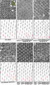
Loss of frl causes a PCP-like defect in the eye. Tangential sections of adult eyes are shown with corresponding schematic representation of ommatidial orientations underneath. Anterior is to the left and dorsal is up in all sections. Black and red arrows represent dorsal and ventral chiral forms of ommatidia, and circles mark lost R-cells in all figures. (A) Control eyes expressing Dcr2 under control of the sevenless (sev) promoter. The inset shows an enlarged single ommatidium with numbered rhabdomeres, the light-sensitive organelles of R-cells (note that only seven rhabdomeres are visible at once in a section, as R7 lies on top of R8). (B) Compared to control (A), knock down of frl causes ommatidial rotation defects. (C–F) frlEx83 (C) and frlEx88 (E) clones marked by the absence of pigment show PCP-like defects (due to the Minute heterozygous background, most of the sections are homozygous mutant). (D and F) Lethality as well as orientation defects of frlEx83 (D) and frlEx88 (F) are rescued by a genomic copy of frl (FosFrl). Note that throughout this work, all alleles of frl correspond to the excision alleles indicated in the background of a FRT80 chromosome with a fosmid adding back all additional genes removed by the excisions (FosAB).
Figure 2
Figure 2
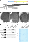
Frl likely is autoinhibited. (A) Schematic of domain structure of Frl with amino acid number of predicted domains indicated (relative to version Frl-PC). GBD, GTPase binding domain; DID, diaphanous inhibitory domain; DD, dimerization domain; FH1 and FH2, formin homology 1/2 domains; DAD, diaphanous autoregulatory domain. Underneath: schematic and coordinates of constructs used in this study; note that FlagΔFH2 is 11 aa shorter than the Gst-ΔFH2. (B–D) Images of eyes of flies overexpressing full-length Frl (B), constitutively active Frl (FrlΔGBD-DID; C), or dominant negative Frl (FrlΔFH2; D) under the control of sev-Gal4. Note that in contrast to full-length Frl that does not affect eye formation, FrlΔGBD-DID and FrlΔFH2 cause severely rough eyes. (E) Gst pull-down experiment of in vitro translated proteins indicated on the left with Gst-fusion proteins indicated on top. While only a small amount of full-length Frl is binding to the FH2-CT domain, much stronger binding is found between the GBD-DID-DD-CC region with Gst-FH2-CT and Gst-CT, consistent with an intramolecular interaction of the N and C termini of Frl. (F) Coomassie staining of a typical pull-down gel of the assays in E with corresponding Gst proteins indicated on top.
Figure 3
Figure 3
Eye phenotypes of constitutively active and dominant negative Frl. (A) Expression of activated sep > CA-Frl (ΔGBD-DID) induces a loss of R-cells. (B) sep > DN-Frl (ΔFH2) causes PCP-like defects reflected mainly by misrotated ommatidia and leads to loss of some R-cells. (C–F) PCP defects caused by sep > FrlΔFH2 arise in third instar eye discs. Confocal images of w1118 (C and E) and sep>ΔFH2 (D and F) third instar eye discs stained for nuclear β-Gal expressed from svp-lacZ07482 enhancer trap line (green in C and D) or stained for Salm marking R3/4 at early stages of rotation (E and F). R-cells are counterstained with ELAV (red). Dorsal parts of the disc just posterior to the MF are shown (top). Schematic of the cluster orientation (bottom).
Figure 4
Figure 4
Frl preferentially binds to Cdc42 in its GTP bound form. (A) Coimmunoprecipitation of the indicated, Myc-tagged constitutively active (Q) or dominant negative (N) human Rho family GTPases by Flag-FrlΔFH2 from HEK293 cell lysates shows that Frl strongly binds Cdc42 Q61L and, as visible on the darker exposure, more weakly to Rac Q61L, but not to their dominant negative versions. Flag-Dsh was used as negative control. (Top) Immunoprecipitations. (Bottom) Cell lysates. (B) In vitro translated [35]S-labeled Frl-GBD-DID-DD-CC binds to purified Gst Drosophila Cdc42 loaded with the nonhydrolyzable GTP analog GMP-PNP, but not to Gst-Cdc42 loaded with GDP or Gst alone. (C) Frl-GBD-DID-DD-CC more weakly binds to purified Gst Drosophila GMP-PNP-Rac1, but only barely to GDP-Rac1 or Gst alone. (D and E) Quantification of typical Cdc42 (D) and Rac1 (E) Gst pull downs performed in triplicates of assays analogous to the ones shown in B and C. t-test; ***P < 0.001; *P < 0.05.
Figure 5
Figure 5
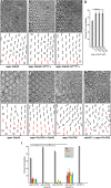
Frl can be activated by Cdc42 in vivo. (A–C) The R-cell loss of sep > Cdc42 (A) is dominantly suppressed by removal of one gene dose of each frl allele (B: frlEx83; C: frlExK62 and not shown; quantification in D). (D) Quantification (percentage ommatidia with wild-type R-cell complement) of sep > Cdc42 lacking one gene dose of the alleles indicated underneath. (E–H) sep > Cdc42 induced R-cell loss (E shows the phenotype of a different chromosomal insertion than used in A) is fully suppressed by coexpression of dominant negative sep > DN-Frl (ΔFH2) (F; quantified in I). Conversely, the phenotype of sep > DN-Frl (G; note that this stronger line also causes gaps in between ommatidia) is suppressed by coexpression of sep > Cdc42 (F), but dominantly enhanced by cdc421, a null allele (H; quantified in I). (I) Quantification of PCP and R-cell loss phenotypes of indicated genotypes (gaps: whole ommatidia missing). t-test; ***P < 0.001; **P < 0.02.
Figure 6
Figure 6
Frl interacts with Dsh in vitro, but not in vivo. (A) Gst pull-down assay of in vitro translated Frl fragments indicated on the left by purified Gst fusion proteins indicated on top. Full-length Frl and Frl-FH2-CT, but not the FH2 domain alone, bind to Gst-Dsh and its basic PDZ region, but not to Gst alone or the Dsh DEP-Cterm domain. The core PCP gene Pk is used as positive control for Dsh binding (Jenny et al. 2005). (Top) Autoradiographs. (Bottom-most) Coomassie stained gel of a typical pull-down showing the Gst proteins. (B) Coimmunoprecipitations of Dsh-GFP by the indicated Myc-tagged Frl fragments from HEK293 cells. Only FrlΔGBD-DID is able to interact with Dsh-GFP. A fragment of the Wnk kinase was used as negative control (Wnk-NT2). (Top) Immunoprecipitations. (Bottom) Cell lysates. (C–E) The PCP phenotype of dsh1 hemizygous males (C) is not altered by the removal of one gene dose of frl (D: frlEx83; E: frlExK62; quantified in F). Tangential eye sections are shown above schematics. (F) Quantification of PCP phenotypes of indicated genotypes.
Figure 7
Figure 7
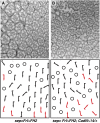
The phenotype of DN-Frl (A) is dominantly enhanced by concurrent removal of one gene dose of both N-cadherins (CadNΔ14; B). Above: tangential eye sections; below: schematics. See Figure S3 for quantification.
Figure 8
Figure 8
Frl cooperates with DAAM during neural development. (A–C) Compared to frl (Figure 1) and DAAMEx68 single (null) mutant R-cells (A), DAAMEx68; frlEx83 (B) and DAAMEx68; frlExK62 (C) double mutant R-cells show a cell-autonomous small rhabdomere phenotype, suggesting that DAAM and Frl have a redundant function in R-cell morphogenesis or maintenance. Double mutant cells lack rhabdomere-associated pigment granules (examples marked with magenta arrowheads), while R-cells of all other genotypes contain pigment (examples marked with blue arrowheads). (D) Schematic of a right part of a mushroom body (MB) of an adult brain with the neuropile lobes growing from the Kenyon cells. Axons of α/β and α′/β′ neurons bifurcate with one axon projecting into a dorsal (α/α′) and one growing into a medial (β/β′) lobe. Magenta frame depicts area shown in confocal images in E–G. (E–G) Positively marked MARCM clones labeled by GFP of wild-type (E), frlEx88 mutant (F), and frlExK62 mutant (G) neurons in the α/β lobes counterstained with Fasciclin II (FasII, red). Wild-type neurons grow axons that terminate (yellow arrowheads in E) at the tip of the α/β lobes (white arrowheads), while axons of frl mutants often arrest prematurely (yellow arrowheads in F and G). (H) Quantification of axons with growth defects (in percentage) of the indicated frl mutant neurons. White numbers indicate number of counted clones. Dorsal is up and medial is to the right in E–G.
Figure 9
Figure 9
Frl and DAAM cooperate in MB axon growth. In all panels, quantifications of defects are shown in percentage for the indicated genotypes with growth defects (shortened, thinner, or absent lobes) in blue and guidance defects (thickened or misprojecting lobes) depicted in red. Numbers indicate MBs scored per genotype (note that α and β lobes were scored in the same samples). (A and B) All three frl alleles dominantly enhance the axon growth defects in α lobes (A) and β lobes (B) of DAAMEx1 and DAAMEx4 hemizygous males. (C and D) Similarly, RNAi-mediated knock down of frlTRIPHMS00445 under control of OK107-Gal4 enhances the axon growth defects of DAAMEx4 in α lobes (C) and DAAMEx1 and DAAMEx4 in β lobes (D). (E and F) The MB phenotype of dsh1 hemizygous males is not altered by removal of a gene dose of frl (E: α lobes; F: β lobes). (G and H) All three frl alleles dominantly enhance the axon growth defects in α lobes (G) and β lobes (H) of cdc422 hemizygous males. Fisher’s exact test; *P < 0.05; **P < 0.005; ***P < 0.0001.

Similar articles

Cited by

References

    1. Abou-Kheir W., Isaac B., Yamaguchi H., Cox D., 2008. Membrane targeting of WAVE2 is not sufficient for WAVE2-dependent actin polymerization: a role for IRSp53 in mediating the interaction between Rac and WAVE2. J. Cell Sci. 121: 379–390. - PMC - PubMed
    1. Axelrod J. D., 2001. Unipolar membrane association of Dishevelled mediates Frizzled planar cell polarity signaling. Genes Dev. 15: 1182–1187. - PMC - PubMed
    1. Basler K., Hafen E., 1990. Receptor tyrosine kinases mediate cell-cell interactions during Drosophila development. Prog. Growth Factor Res. 2: 15–27. - PubMed
    1. Basler K., Christen B., Hafen E., 1991. Ligand-independent activation of the sevenless receptor tyrosine kinase changes the fate of cells in the developing Drosophila eye. Cell 64: 1069–1081. - PubMed
    1. Block J., Breitsprecher D., Kuhn S., Winterhoff M., Kage F., et al. , 2012. FMNL2 drives actin-based protrusion and migration downstream of Cdc42. Curr. Biol. 22: 1005–1012. - PMC - PubMed

Publication types

MeSH terms