Review
. 2016 Aug 15;594(16):4591-613.
doi: 10.1113/JP271471. Epub 2016 Mar 17.

Physiological and pathophysiological reactive oxygen species as probed by EPR spectroscopy: the underutilized research window on muscle ageing

Affiliations
Review

Physiological and pathophysiological reactive oxygen species as probed by EPR spectroscopy: the underutilized research window on muscle ageing

Engy A Abdel-Rahman et al. J Physiol. .

Abstract

Reactive oxygen and nitrogen species (ROS and RNS) play crucial roles in triggering, mediating and regulating physiological and pathophysiological signal transduction pathways within the cell. Within the cell, ROS efflux is firmly controlled both spatially and temporally, making the study of ROS dynamics a challenging task. Different approaches have been developed for ROS assessment; however, many of these assays are not capable of direct identification or determination of subcellular localization of different ROS. Here we highlight electron paramagnetic resonance (EPR) spectroscopy as a powerful technique that is uniquely capable of addressing questions on ROS dynamics in different biological specimens and cellular compartments. Due to their critical importance in muscle functions and dysfunction, we discuss in some detail spin trapping of various ROS and focus on EPR detection of nitric oxide before highlighting how EPR can be utilized to probe biophysical characteristics of the environment surrounding a given stable radical. Despite the demonstrated ability of EPR spectroscopy to provide unique information on the identity, quantity, dynamics and environment of radical species, its applications in the field of muscle physiology, fatiguing and ageing are disproportionately infrequent. While reviewing the limited examples of successful EPR applications in muscle biology we conclude that the field would greatly benefit from more studies exploring ROS sources and kinetics by spin trapping, protein dynamics by site-directed spin labelling, and membrane dynamics and global redox changes by spin probing EPR approaches.


Figures

Figure 1
Figure 1
Mitochondrial ROS generation and cascade of transformations due to interaction with cellular antioxidants and metals
Figure 2
Figure 2. Estimation of spin concentration of DMPO‐OH spin adduct using standard TEMPOL calibration
A, linear dependence of the EPR signal's amplitude on TEMPOL concentration. B, calibration using double integration to obtain spin concentration also yielded linear relationship with TEMPOL concentration. C, kinetics of hydroxyl radical production through Fe2+ + H2O2 Fenton chemistry in the presence of DMPO in PBS buffer adjusted to pH = 7.4 at 37°C. Final concentrations were: 0.1 mm FeSO4, 200 mm DMPO, 1 mm H2O2. EPR parameters were: centre field, 3338.26; sweep width, 0.0001 mT; sweep time, 22 min; modulation amplitude, ±0.2 mT; microwave frequency, 9.75 GHz.
Figure 3
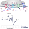
Figure 3. EPR‐detected mitochondrial iron‐sulfur centres in frozen mouse liver
Freshly extracted mouse liver is pulverized and loaded in an EPR quartz tube. The EPR tube was then slowly fitted in a glass Dewar flask filled with liquid nitrogen at equilibrium inside a Magnettech Benchtop EPR spectrometer before starting spectral acquisition. The resulting EPR spectrum reflects iron‐sulfur centres of the partially reduced mitochondrial respiratory chain complexes I–III (upper panel) and are assigned as described in van der Kraaij et al. (1989). EPR parameters: sweep rate, 1.55 mT s−1; modulation amplitude, 0.7 mT; MW attenuation, 10 dB; gain, 200.
Figure 4
Figure 4. Pictorial depiction of the ability of EPR technique to detect ROS and probe redox changes in cellular compartments including membranes
Spin traps DEPMPO (distributed in extra‐ and intracellular spaces) and DIPPMPO (can also localize in membranes) can be used to trap superoxide, hydroxyl, carbon‐centred and sulfur‐centred radicals in various cellular locations. Spin labels N‐(1‐Oxyl‐2,2,6,6‐tetramethyl‐4‐piperidinyl)maleimide (MAL‐6) or (1‐Oxyl‐2,2,5,5‐tetramethyl‐Δ3‐pyrroline‐3‐methyl) Methanethiosulfonate (MTSSL) specifically target free thiol bonds in cysteine residues and would therefore reveal information on conformational changes due to S–S bond formation. Analyses of the EPR lineshape and intensity of doxyl stearic acids 5‐DSA and 16‐DSA at conditions exhibiting variable oxidative stress level would provide information on the lipid peroxidation, ROS and oxygen flux within phospholipid bilayers as well as on membrane fluidity. Spin‐labelled doxyl stearic acids have also been used to distinguish various membrane regions with different fluidity characteristics while probing protein dynamics.
Figure 5
Figure 5. Analysis of the stereochemistry of addition of superoxide radical onto DEPMPO may reveal information on the environment where spin trapping occurs
Encounter in a fluid environment is expected to yield both cis and trans diastereoisomeric nitroxides with near equal proportions (A); a case observed when inhibition of complex III with antimycin A was carried out (B). Restricted environments would favour one of the diastereoisomers as seen in the case of complex I inhibition by rotenone (C). Spectral simulations were carried out to estimate the type and contributions of each radical adduct using WinSim software and reported EPR parameters for both cis and trans adducts with DEPMPO (Culcasi et al. 2006).
Figure 6
Figure 6. Probing membrane properties by spin‐labelling EPR spectroscopy
Spin‐label nitroxide ring is rigidly attached to the 5th carbon (column A, 5‐DSA, probes membrane rigidity close to the hydrophilic surface of the membrane) or the 16th carbon (column B, 16‐DSA, probes the fluidity in the core of the phospholipid bilayer). When in non‐viscous media, both spin labels exhibit sharp EPR spectral peaks, but upon their insertion into lipid bilayers these spin labels undergo dramatic spectral changes due to restricted motion of the spin labels. This is mainly because hyperfine coupling is dependent on the relative orientation of the molecular orbital where the free electron resides and on the applied external magnetic field. In disordered fluid phase (non‐viscous solvents) the fast reorientational motion of the spin label leads to isotropic hyperfine coupling. However, in the ordered membrane phase, reorientation is not hindered by the surrounding ordered molecules leading to anisotropic hyperfine interaction, which is reflected in asymmetric EPR lines. In some cases, EPR spectra of ordered spin labels show more than one component reflecting regional variability in membrane fluidity due, for example, to being in cholesterol‐rich versus ‐poor membrane domains. Lineshape analyses can yield order parameter, rotational correlation time and relative proportions of rigid (R) versus fluid (F) regions.

Similar articles

Cited by

References

    1. Abbas K, Hardy M, Poulhes F, Karoui H, Tordo P, Ouari O & Peyrot F (2014). Detection of superoxide production in stimulated and unstimulated living cells using new cyclic nitrone spin traps. Free Radic Biol Med 71, 281–290. - PubMed
    1. Akaike T & Maeda H (1996). Quantitation of nitric oxide using 2‐phenyl‐4,4,5,5‐tetramethylimidazoline‐1‐oxyl 3‐oxide (PTIO). Methods Enzymol 268, 211–221. - PubMed
    1. Ali SS, Hsiao M, Zhao HW, Dugan LL, Haddad GG & Zhou D (2012). Hypoxia‐adaptation involves mitochondrial metabolic depression and decreased ROS leakage. PLoS One 7, e36801. - PMC - PubMed
    1. Ali SS, Marcondes MC, Bajova H, Dugan LL & Conti B (2010). Metabolic depression and increased reactive oxygen species production by isolated mitochondria at moderately lower temperatures. J Biol Chem 285, 32522–32528. - PMC - PubMed
    1. Ali SS, Xiong C, Lucero J, Behrens MM, Dugan LL & Quick KL (2006). Gender differences in free radical homeostasis during aging: shorter‐lived female C57BL6 mice have increased oxidative stress. Aging Cell 5, 565–574. - PubMed

Publication types

Substances

LinkOut - more resources