. 2016 Apr 1;310(7):H873-90.
doi: 10.1152/ajpheart.00682.2015. Epub 2016 Jan 22.

Myocyte repolarization modulates myocardial function in aging dogs

Affiliations

Myocyte repolarization modulates myocardial function in aging dogs

Andrea Sorrentino et al. Am J Physiol Heart Circ Physiol. .

Abstract

Studies of myocardial aging are complex and the mechanisms involved in the deterioration of ventricular performance and decreased functional reserve of the old heart remain to be properly defined. We have studied a colony of beagle dogs from 3 to 14 yr of age kept under a highly regulated environment to define the effects of aging on the myocardium. Ventricular, myocardial, and myocyte function, together with anatomical and structural properties of the organ and cardiomyocytes, were evaluated. Ventricular hypertrophy was not observed with aging and the structural composition of the myocardium was modestly affected. Alterations in the myocyte compartment were identified in aged dogs, and these factors negatively interfere with the contractile reserve typical of the young heart. The duration of the action potential is prolonged in old cardiomyocytes contributing to the slower electrical recovery of the myocardium. Also, the remodeled repolarization of cardiomyocytes with aging provides inotropic support to the senescent muscle but compromises its contractile reserve, rendering the old heart ineffective under conditions of high hemodynamic demand. The defects in the electrical and mechanical properties of cardiomyocytes with aging suggest that this cell population is an important determinant of the cardiac senescent phenotype. Collectively, the delayed electrical repolarization of aging cardiomyocytes may be viewed as a critical variable of the aging myopathy and its propensity to evolve into ventricular decompensation under stressful conditions.

Keywords: aging; contractile reserve; myocardium.


Figures

Fig. 1.
Fig. 1.
Age of adult (A) and old (O) dogs employed in various tests is shown as median and interquartile ranges. References to figure panels are reported.
Fig. 2.
Fig. 2.
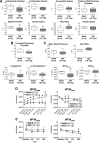
Aging negatively interferes with cardiac function. A: echocardiographic parameters in aging dogs; data are shown as median and interquartile ranges (adult: n = 6, 5.5 ± 0.6 yr; old: n = 8, 11.9 ± 0.4 yr). *P < 0.05 vs. adult; y, yr of age. B: radial left ventricular (LV) contractility in aging dogs; data are shown as median and interquartile ranges (adult: n = 5, 5.9 ± 0.6 yr; old: n = 8, 11.8 ± 0.4 yr). *P < 0.05 vs. adult. C: hemodynamic parameters in aging dogs; data are shown as median and interquartile ranges (adult: n = 6, 5.5 ± 0.6 yr; old: n = 9, 12.0 ± 0.4 yr). *P < 0.05 vs. adult. D: maximal rate of LV contraction and relaxation in aging dogs following infusion of dobutamine (Dobt, dosage referred to kg of body wt per minute of infusion) and bolus of isoproterenol (Iso, dosage referred to kg of body wt) are shown as mean ± SE (adult: n = 6, 5.5 ± 0.6 yr; old: n = 6, 12.5 ± 0.4 yr). *P < 0.05 vs. adult; †P < 0.001 within the same group E: effects of overdrive pacing are shown as mean ± SE (adult: n = 6, 5.5 ± 0.6 yr; old: n = 5 12.0 ± 0.6 yr). *P < 0.05 vs. adult; †P < 0.01 within the same group. F: ECGs from precordial leads obtained in a dog at 11 yr of age. Notched R waves are present in V6. Arrow points to the notched R wave in the magnified trace. G: gross anatomical parameters in aging dogs. Body weight before death and heart weight and LV free wall thickness measured in the explanted organ (male: n = 21; female: n = 18). Data are fitted with linear regression. R2 and P value for each fitting are reported. H: biochemical parameters of blood samples obtained from the jugular vein, with the exception of lactate and partial pressure of oxygen, measured in blood collected from the coronary sinus. Data are fitted with linear regression. R2 and P values for each fitting are reported. I–L: Western blots analysis for total and phosphorylated PKA and phospholamban (PLB) in the LV myocardium of aging dogs. Quantitative data for p-PKA/PKA ratio (J) in the LV myocardium of adult (n = 11, 5.2 ± 0.4 yr) and old (n = 12, 12.9 ± 0.2 yr) beagle dogs are shown as median and interquartile ranges. Quantitative data for p-PLB/PLB ratio (L) in the LV myocardium of adult (n = 10, 5.2 ± 0.5 yr) and old (n = 12, 12.9 ± 0.2 yr) beagle dogs are shown as median and interquartile ranges.
Fig. 2.
Fig. 2.
Aging negatively interferes with cardiac function. A: echocardiographic parameters in aging dogs; data are shown as median and interquartile ranges (adult: n = 6, 5.5 ± 0.6 yr; old: n = 8, 11.9 ± 0.4 yr). *P < 0.05 vs. adult; y, yr of age. B: radial left ventricular (LV) contractility in aging dogs; data are shown as median and interquartile ranges (adult: n = 5, 5.9 ± 0.6 yr; old: n = 8, 11.8 ± 0.4 yr). *P < 0.05 vs. adult. C: hemodynamic parameters in aging dogs; data are shown as median and interquartile ranges (adult: n = 6, 5.5 ± 0.6 yr; old: n = 9, 12.0 ± 0.4 yr). *P < 0.05 vs. adult. D: maximal rate of LV contraction and relaxation in aging dogs following infusion of dobutamine (Dobt, dosage referred to kg of body wt per minute of infusion) and bolus of isoproterenol (Iso, dosage referred to kg of body wt) are shown as mean ± SE (adult: n = 6, 5.5 ± 0.6 yr; old: n = 6, 12.5 ± 0.4 yr). *P < 0.05 vs. adult; †P < 0.001 within the same group E: effects of overdrive pacing are shown as mean ± SE (adult: n = 6, 5.5 ± 0.6 yr; old: n = 5 12.0 ± 0.6 yr). *P < 0.05 vs. adult; †P < 0.01 within the same group. F: ECGs from precordial leads obtained in a dog at 11 yr of age. Notched R waves are present in V6. Arrow points to the notched R wave in the magnified trace. G: gross anatomical parameters in aging dogs. Body weight before death and heart weight and LV free wall thickness measured in the explanted organ (male: n = 21; female: n = 18). Data are fitted with linear regression. R2 and P value for each fitting are reported. H: biochemical parameters of blood samples obtained from the jugular vein, with the exception of lactate and partial pressure of oxygen, measured in blood collected from the coronary sinus. Data are fitted with linear regression. R2 and P values for each fitting are reported. I–L: Western blots analysis for total and phosphorylated PKA and phospholamban (PLB) in the LV myocardium of aging dogs. Quantitative data for p-PKA/PKA ratio (J) in the LV myocardium of adult (n = 11, 5.2 ± 0.4 yr) and old (n = 12, 12.9 ± 0.2 yr) beagle dogs are shown as median and interquartile ranges. Quantitative data for p-PLB/PLB ratio (L) in the LV myocardium of adult (n = 10, 5.2 ± 0.5 yr) and old (n = 12, 12.9 ± 0.2 yr) beagle dogs are shown as median and interquartile ranges.
Fig. 3.
Fig. 3.
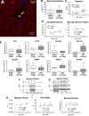
Structural properties of the myocardium are modestly affected by aging in beagle dogs. A: apoptotic myocyte (arrow) in the adult canine myocardium by terminal deoxynucleotidyl transferase-mediated dUTP nick-end labeling (TUNEL; TdT; green) assay. Myocytes are identified by α-sarcomeric actin (α-SA; red). Nuclei are stained by DAPI (blue). B: quantitative data for myocyte apoptosis in the LV myocardium of adult (n = 5, 4.8 ± 0.4 yr) and old (n = 6, 12.9 ± 0.3 yr) beagle dogs are shown as median and interquartile ranges. C: circulating levels of cardiac troponin I in beagle dogs (n = 63). Data are fitted with linear regression. R2 and P values are reported. D: quantitative measurements of interstitial fibrosis (n = 23) and volume fraction (n = 22) of cardiomyocytes. Data are fitted with linear regression. R2 and P values for each fitting are reported. E: quantitative data for level of transcript for fibronectin (FN1), connective tissue growth factor (CTGF), collagen type I α1 (COL1A1), collagen type I α2 (COL1A2), collagen type III α1 (COL3A1), matrix metalloproteinase-II (MMP2), transforming growth factor-β1 (TGFB1), and periostin (POSTN) in the LV of adult and old beagle dogs. Data are shown as median and interquartile ranges (adult: n = 6 specimens, 6 dogs, 4.4 ± 0.4 yr; old: n = 6 specimens, 6 dogs, 13.1 ± 0.2 yr). *P < 0.05 vs. adult. F: volume of cardiomyocytes obtained from beagle dogs at 4, 7–8 and 12–14 yr of age (n = 404 myocytes, 3 dogs, 3.8 ± 0.03 yr; n = 309 myocytes, 4 dogs, 7.9 ± 0.2 yr; n = 360 myocytes, 4 dogs, 13.1 ± 0.2 yr, respectively) is shown as median and interquartile ranges. *P < 0.01 vs. 4 yr. G: fraction of mononucleated and binucleated myocytes for cells shown in F. H: quantitative data for myocyte volume shown in F are reported as mean ± SE and fitted with linear regression. R2 and P values are reported. I: myocardial mass and computed myocyte number in aging beagle dogs (n = 38). Data are fitted with linear regression. R2 and P values for each fitting are reported.
Fig. 4.
Fig. 4.
Aging results in prolongation of the electrical recovery of myocardium and myocytes. A: pseudo-ECGs in the perfused adult and old LV myocardium stimulated at 2 Hz. Superimposed traces are shown in the inset. B: quantitative measurements of QT interval duration in the myocardium of beagle dogs (n = 19 muscles, 19 dogs) are fitted with linear regression. R2 and P values are reported. C: monophasic action potentials (MAPs) measured in adult and old LV tissue. D: quantitative measurements of duration of the action potential (AP) at 90% repolarization (APD90; n as in B) are fitted with linear regression. R2 and P values are reported. E: superimposed pseudo-ECGs obtained at different pacing cycle lengths (CL). F: data are shown as mean ± SE (adult: n = 11 muscles, 6 dogs, 3.8 ± 0.4 yr; old: n = 8 muscles, 7 dogs, 11.5 ± 0.4 yr). *P < 0.05 vs. adult; †P < 0.001 for various frequencies within the same group. G: APs of LV myocytes from adult and old dogs. H: data are shown as mean ± SE (adult: n = 22 cells, 8 dogs, 5.6 ± 0.4 yr; old: n = 10 cells, 5 dogs, 13.1 ± 0.1 yr). *P < 0.05 vs. adult. RMP, resting membrane potential; APA, AP amplitude. I: superimposed APs at various pacing frequencies in a LV myocyte from an adult dog. J: data are shown as mean ± SE (adult: n = 16 cells, 2 dogs, 3.2 ± 0 yr; old: n = 26 cells, 6 dogs, 10.9 ± 0.1 yr). *P < 0.05 vs. adult; †P < 0.001 for various frequencies within the same group.
Fig. 4.
Fig. 4.
Aging results in prolongation of the electrical recovery of myocardium and myocytes. A: pseudo-ECGs in the perfused adult and old LV myocardium stimulated at 2 Hz. Superimposed traces are shown in the inset. B: quantitative measurements of QT interval duration in the myocardium of beagle dogs (n = 19 muscles, 19 dogs) are fitted with linear regression. R2 and P values are reported. C: monophasic action potentials (MAPs) measured in adult and old LV tissue. D: quantitative measurements of duration of the action potential (AP) at 90% repolarization (APD90; n as in B) are fitted with linear regression. R2 and P values are reported. E: superimposed pseudo-ECGs obtained at different pacing cycle lengths (CL). F: data are shown as mean ± SE (adult: n = 11 muscles, 6 dogs, 3.8 ± 0.4 yr; old: n = 8 muscles, 7 dogs, 11.5 ± 0.4 yr). *P < 0.05 vs. adult; †P < 0.001 for various frequencies within the same group. G: APs of LV myocytes from adult and old dogs. H: data are shown as mean ± SE (adult: n = 22 cells, 8 dogs, 5.6 ± 0.4 yr; old: n = 10 cells, 5 dogs, 13.1 ± 0.1 yr). *P < 0.05 vs. adult. RMP, resting membrane potential; APA, AP amplitude. I: superimposed APs at various pacing frequencies in a LV myocyte from an adult dog. J: data are shown as mean ± SE (adult: n = 16 cells, 2 dogs, 3.2 ± 0 yr; old: n = 26 cells, 6 dogs, 10.9 ± 0.1 yr). *P < 0.05 vs. adult; †P < 0.001 for various frequencies within the same group.
Fig. 5.
Fig. 5.
A: APs recorded in 1 adult and 1 old myocyte in Tyrode (Tyr) and after exposure to mexiletine (Mex; 10 μM). B: data are shown as mean ± SE (adult: n = 19 cells, 8 dogs, 5.2 ± 0.4 yr; old: n = 10 cells, 4 dogs, 13.1 ± 0.1 yr). *P < 0.001 vs. Tyr. C: comparison of the effects of late Na+ current (INaL) inhibition on AP duration between adult (A) and old (O) myocytes reported in B. data are shown as mean ± SE. *P < 0.01 vs. A. D: pseudo-ECG in the perfused LV myocardium of an adult dog. Traces in Krebs-Henseleit buffer (KHB) solution and after exposure to Mex (10 μM) are reported and superimposed in inset. E: data are shown as mean ± SE (adult: n = 6 muscles, 6 dogs, 6.5 ± 0.9 yr; old: n = 6 muscles, 4 dogs, 12.7 ± 0.5 yr). *P < 0.05 vs. KHB. F: superimposed traces of INaL (top traces) measured in voltage-clamp using a voltage-command protocol (bottom trace) at 0.5, 1.5, and 3 Hz, in a juvenile hound type dog myocyte. INaL was progressively reduced at higher frequencies. G: quantitative data for INaL reactivation in LV myocytes form juvenile (0.6–0.8 year old) hound type dogs are reported as mean ± SE (n = 29 cells, 4 dogs). †P < 0.001 for various frequencies. Amplitude of INaL is reported as positive value. H and I: data relative to effects of INaL inhibitors ranolazine (Ran; 10 μM) (H) and low doses of tetrodotoxin (TTX; 1 μM) (I) on AP profile of cardiomyocytes from old beagle dogs (11–13 yr of age) are shown as mean ± SE (ranolazine: n = 10 cells, 2 dogs 11 yr old; TTX: n = 5 cells, 2 dogs, 11–13 yr). *P < 0.05 vs. Tyr; †P < 0.001 for various frequencies within the same group.
Fig. 5.
Fig. 5.
A: APs recorded in 1 adult and 1 old myocyte in Tyrode (Tyr) and after exposure to mexiletine (Mex; 10 μM). B: data are shown as mean ± SE (adult: n = 19 cells, 8 dogs, 5.2 ± 0.4 yr; old: n = 10 cells, 4 dogs, 13.1 ± 0.1 yr). *P < 0.001 vs. Tyr. C: comparison of the effects of late Na+ current (INaL) inhibition on AP duration between adult (A) and old (O) myocytes reported in B. data are shown as mean ± SE. *P < 0.01 vs. A. D: pseudo-ECG in the perfused LV myocardium of an adult dog. Traces in Krebs-Henseleit buffer (KHB) solution and after exposure to Mex (10 μM) are reported and superimposed in inset. E: data are shown as mean ± SE (adult: n = 6 muscles, 6 dogs, 6.5 ± 0.9 yr; old: n = 6 muscles, 4 dogs, 12.7 ± 0.5 yr). *P < 0.05 vs. KHB. F: superimposed traces of INaL (top traces) measured in voltage-clamp using a voltage-command protocol (bottom trace) at 0.5, 1.5, and 3 Hz, in a juvenile hound type dog myocyte. INaL was progressively reduced at higher frequencies. G: quantitative data for INaL reactivation in LV myocytes form juvenile (0.6–0.8 year old) hound type dogs are reported as mean ± SE (n = 29 cells, 4 dogs). †P < 0.001 for various frequencies. Amplitude of INaL is reported as positive value. H and I: data relative to effects of INaL inhibitors ranolazine (Ran; 10 μM) (H) and low doses of tetrodotoxin (TTX; 1 μM) (I) on AP profile of cardiomyocytes from old beagle dogs (11–13 yr of age) are shown as mean ± SE (ranolazine: n = 10 cells, 2 dogs 11 yr old; TTX: n = 5 cells, 2 dogs, 11–13 yr). *P < 0.05 vs. Tyr; †P < 0.001 for various frequencies within the same group.
Fig. 6.
Fig. 6.
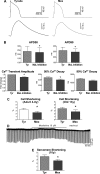
INaL has inotropic effects on cardiomyocytes. A: AP (top traces) and Ca2+ transients (bottom traces) recorded simultaneously in a myocyte from an old dog in Tyr and after exposure to the INaL inhibitor Mex (10 μM). B: quantitative data for AP and Ca2+ transient properties in old myocytes, in Tyr, and in the presence of INaL inhibition (10 μM Mex, n = 1, or 1 μM TTX, n = 3); data are shown as mean ± SE (n = 4 cells, 2 dogs, 11–14 yr old). *P < 0.05 vs. Tyr. C: data on contractile (cell shortening) properties in adult (left: n = 14 cells, 6 dogs, 6.3 ± 0.9 yr) and old (right: n = 6 cells, 2 dogs, 13.1 ± 0.3 yr) myocytes stimulated at 0.25–0.5 Hz in Tyr and following exposure to Mex (10 μM) are shown as mean ± SE. *P < 0.05. D: trace of sarcomere shortening for an old myocyte during the exposure to Mex and during the washout phase. E: data of contractility (sarcomere shortening) for old myocytes are shown as mean ± SE (n = 7 cells, 1 dog, 10 yr old). *P < 0.001 vs. Tyr.
Fig. 7.
Fig. 7.
The old myocardium presents blunted contractile force frequency relationship. A: isometric contraction of ventricular trabeculae obtained from adult and old dogs stimulated at 1-Hz pacing frequency. Twitches are superimposed in the inset. B: data are shown as mean ± SE (adult: n = 9 muscles, 6 dogs, 5.8 ± 0.6 yr; old: n = 10 muscles, 7 dogs, 13.2 ± 0.1 yr). *P < 0.05. C: isometric contraction (top traces) and first derivative of developed tension (bottom traces) recorded at progressively higher stimulation frequencies in adult and old trabeculae. D and E: quantitative data for developed tension (D) and time to 90% relaxation (E) normalized with respect to CL 2,000 ms are shown as mean ± SE (adult: n = 7 muscles, 5 dogs, 5.1 ± 0.6 yr; old: n = 6 muscles, 4 dogs, 12.2 ± 0.4 yr). *P < 0.05 vs. adult; †P < 0.001 for various frequencies within the same group.
Fig. 8.
Fig. 8.
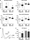
The old myocardium presents blunted contractile response to β- adrenergic receptor (β-AR) stimulation. A: data for twitch properties in aging muscles in KHB and after exposure to isoproterenol (Iso; 100 nM) are shown as median and interquartile ranges (adult: n = 9 muscles, 7 dogs, 5.5 ± 0.5 yr; old: n = 10 muscles, 8 dogs, 13.0 ± 0.2 yr). *P < 0.01 vs. KHB. B: superimposed pseudo-ECGs obtained in the perfused adult LV myocardium in KHB and after exposure to the β-AR agonist Iso (100 nM). C: quantitative data for QT interval in adult and old LV myocardium in KHB and after exposure to Iso are shown as mean ± SE (adult: n = 4 muscles, 4 dogs, 6.9 ± 1 yr; old: n = 7 muscles, 5 dogs, 12.8 ± 0.3 yr). *P < 0.05 vs. KHB.
Fig. 9.
Fig. 9.
INaL alters mechanical properties of the ventricular myocardium. A: isometric contraction (top trace) and first derivative of developed tension (bottom trace) recorded in a trabecula from an old dog before and after exposure to Mex (10 μM). Twitches and first derivative of developed tension are superimposed in inset. B: data are shown as mean ± SE (adult: n = 6 muscles, 5 dogs, 5.0 ± 0.6 yr; old: n = 10 muscles, 7 dogs, 12.6 ± 0.4 yr). *P < 0.01 vs. KHB. C: isometric contraction (top trace) and first derivative of developed tension (bottom trace) recorded in a trabecula from an old dog before and after exposure to low dose of TTX (1 μM). D: data for developed tension in 3 trabeculae before and after exposure to TTX (1 μM) or Ran (10 μM). Trabeculae were obtained from 2 old dogs (11.3 and 14.3 yr old). E: data for developed tension, normalized with respect to CL 2,000 ms, are shown as mean ± SE (adult: n = 5 muscles, 4 dogs, 5.3 ± 0.6 yr; old: n = 4 muscles, 3 dogs, 12.3 ± 0.7 yr). *P < 0.05 vs. KHB; †P < 0.01 for various frequencies within the same group.
Fig. 10.
Fig. 10.
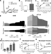
INaL influences the contractile force frequency relationship. A: superimposed pseudo-ECGs obtained in the perfused adult LV myocardium before (KHB) and after exposure to the INaL enhancer ATX-II (5 nM). Arrow points to notched T wave occurring after prolonged exposure to ATX-II. B: data obtained in adult dogs are shown as mean ± SE (n = 4 muscles, 4 dogs, 4–8 yr old). *P < 0.05. Notched T waves were present in 50% of muscles exposed to ATX-II. C: examples of QT interval duration at various stimulation frequencies for LV myocardium obtained from two adult dogs (6 and 7 yr old) in KHB and after exposure to ATX-II. D: isometric contraction (top traces) and first derivative of developed tension (bottom traces) recorded at progressively higher stimulation frequencies in 1 trabecula from an adult dog before (KHB) and after exposure to ATX-II (10 nM). E: data for the effects of ATX-II on developed tension at the various frequencies of stimulation, with respect to baseline, are reported as mean ± SE (n = 3 muscles, 2 dogs, 6 and 7 yr old). *P < 0.05 vs. 333 ms; **P < 0.05 vs. 500 ms; ***P < 0.05 vs. 1,000 ms. F and G: quantitative data for developed tension (F) and developed tension normalized with respect to CL 2,000 ms (G) in adult trabeculae presented in E are shown as mean ± SE. *P < 0.05 vs. KHB; †P < 0.01 for various frequencies within the same group.

Similar articles

Cited by

References

    1. Aiba T, Barth AS, Hesketh GG, Hashambhoy YL, Chakir K, Tunin RS, Greenstein JL, Winslow RL, Kass DA, Tomaselli GF. Cardiac resynchronization therapy improves altered Na channel gating in canine model of dyssynchronous heart failure. Circ Arrhythm Electrophysiol 6: 546–554, 2013. - PMC - PubMed
    1. Aiba T, Hesketh GG, Barth AS, Liu T, Daya S, Chakir K, Dimaano VL, Abraham TP, O'Rourke B, Akar FG, Kass DA, Tomaselli GF. Electrophysiological consequences of dyssynchronous heart failure and its restoration by resynchronization therapy. Circulation 119: 1220–1230, 2009. - PMC - PubMed
    1. Belardinelli L, Giles WR, Rajamani S, Karagueuzian HS, Shryock JC. Cardiac late Na(+) current: proarrhythmic effects, roles in long QT syndromes, and pathological relationship to CaMKII and oxidative stress. Heart Rhythm 12: 440–448, 2015. - PubMed
    1. Bernstein RD, Zhang X, Zhao G, Forfia P, Tuzman J, Ochoa F, Ochoa M, Vogel T, Hintze TH. Mechanisms of nitrate accumulation in plasma during pacing-induced heart failure in conscious dogs. Nitric Oxide 1: 386–396, 1997. - PubMed
    1. Boyle AJ, Shih H, Hwang J, Ye J, Lee B, Zhang Y, Kwon D, Jun K, Zheng D, Sievers R, Angeli F, Yeghiazarians Y, Lee R. Cardiomyopathy of aging in the mammalian heart is characterized by myocardial hypertrophy, fibrosis and a predisposition towards cardiomyocyte apoptosis and autophagy. Exp Gerontol 46: 549–559, 2011. - PMC - PubMed

Publication types

LinkOut - more resources