. 2016 Mar 18;291(12):6483-93.
doi: 10.1074/jbc.M115.691352. Epub 2016 Jan 22.

MicroRNA-7 Regulates the Function of Mitochondrial Permeability Transition Pore by Targeting VDAC1 Expression

Affiliations

MicroRNA-7 Regulates the Function of Mitochondrial Permeability Transition Pore by Targeting VDAC1 Expression

Amrita Datta Chaudhuri et al. J Biol Chem. .

Abstract

Mitochondrial dysfunction is one of the major contributors to neurodegenerative disorders including Parkinson disease. The mitochondrial permeability transition pore is a protein complex located on the mitochondrial membrane. Under cellular stress, the pore opens, increasing the release of pro-apoptotic proteins, and ultimately resulting in cell death. MicroRNA-7 (miR-7) is a small non-coding RNA that has been found to exhibit a protective role in the cellular models of Parkinson disease. In the present study, miR-7 was predicted to regulate the function of mitochondria, according to gene ontology analysis of proteins that are down-regulated by miR-7. Indeed, miR-7 overexpression inhibited mitochondrial fragmentation, mitochondrial depolarization, cytochrome c release, reactive oxygen species generation, and release of mitochondrial calcium in response to 1-methyl-4-phenylpyridinium (MPP(+)) in human neuroblastoma SH-SY5Y cells. In addition, several of these findings were confirmed in mouse primary neurons. Among the mitochondrial proteins identified by gene ontology analysis, the expression of voltage-dependent anion channel 1 (VDAC1), a constituent of the mitochondrial permeability transition pore, was down-regulated by miR-7 through targeting 3'-untranslated region of VDAC1 mRNA. Similar to miR-7 overexpression, knockdown of VDAC1 also led to a decrease in intracellular reactive oxygen species generation and subsequent cellular protection against MPP(+). Notably, overexpression of VDAC1 without the 3'-UTR significantly abolished the protective effects of miR-7 against MPP(+)-induced cytotoxicity and mitochondrial dysfunction, suggesting that the protective effect of miR-7 is partly exerted through promoting mitochondrial function by targeting VDAC1 expression. These findings point to a novel mechanism by which miR-7 accomplishes neuroprotection by improving mitochondrial health.

Keywords: 1-methyl-4-phenylpyridinium; Parkinson disease; microRNA (miRNA); mitochondrial membrane potential; mitochondrial permeability transition (MPT); voltage-dependent anion channel (VDAC).


Figures

FIGURE 1.
FIGURE 1.
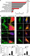
miR-7 regulates mitochondrial function. A, gene ontology analysis of proteins significantly down-regulated by miR-7. Mitochondrial proteins were significantly enriched. GO terms related with mitochondria are indicated in red. B and C, miR-7 inhibits the fragmentation/clumping of mitochondria in response to MPP+. SH-SY5Y cells (B) or primary mouse cortical neurons (C) were transduced with lenti-miR-SC or lenti-miR-7 (red). Three days after transduction, cells were treated with 2 mm (SH-SY5Y) or 0.5 mm MPP+ (primary neurons) for 12 h. Cells were stained with anti-TOM20 (green). D and E, quantification of mitochondrial fragmentation (clumping) was performed in SH-SY5Y (D) and primary neurons (E). These results (B–E) are representatives of three sets of independent experiments. *, p < 0.05, **, p < 0.01.
FIGURE 2.
FIGURE 2.
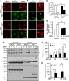
miR-7 regulates the function of mitochondrial PTP. A–D, miR-7 prevents depolarization of mitochondria in response to MPP+. A, SH-SY5Y cells were transfected with miR-SC or miR-7. Forty-eight hours after transfection, cells were treated with 2 mm MPP+ for 12 h. Cells were labeled with JC-1 and imaged. Upon exposure to MPP+, mitochondria of cells transfected with miR-SC were depolarized (green). B, primary mouse neurons were transduced with lenti-miR-SC or lenti-miR-7 (red). Three days after transduction, cells were treated with 0.5 mm MPP+ for 12 h. Upon exposure to MPP+, mitochondria of cells transduced with lenti-miR-SC were depolarized (green). Red fluorescence indicates the lentiviral transduction. C, quantification of JC-1 fluorescence intensities from A. The ratio of red (healthy mitochondria) to green (depolarized mitochondria) fluorescent intensities was calculated from five different microscopic fields for each sample. D, quantification of JC-1 fluorescence intensities from B. Green (depolarized mitochondria) fluorescent intensity was measured from at least 10 transduced (RFP-positive) neurons for each sample. These results (A–D) are representatives of three sets of independent experiments. E, miR-7 prevents release of pro-apoptotic proteins from mitochondria to cytosol. SH-SY5Y cells were transduced with lenti-miR-SC or lenti-miR-7. Three days after transduction, cells were treated with 0.5 mm MPP+ for 6 or 12 h. Cells were lysed, and mitochondrial and cytosolic fractions were isolated. Western blotting analysis was performed. The arrow denotes the position of cytochrome c (Cyt C). This result is a representative of two sets of independent experiments. F and G, miR-7 lowers ROS level in response to MPP+ in SH-SY5Y cells (F) and primary mouse cortical neurons (G). Forty-eight hours after transfection, cells were treated with the indicated concentrations of MPP+ for 12 h in SH-SY5Y cells. Primary mouse neurons were transduced with lenti-miR-SC or lenti-miR-7. Three days after transduction, cells were treated with 0.5 mm MPP+ for 12 h. Data are shown as the means ± S.E. *, p < 0.05, ***, p < 0.005. These results (F and G) are representatives of three sets of independent experiments performed in triplicates for each group.
FIGURE 3.
FIGURE 3.
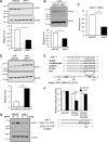
miR-7 down-regulates the mitochondrial PTP component VDAC1. A and B, miR-7 down-regulates VDAC1 protein expression. SH-SY5Y cells (A) were transfected with miR-SC or miR-7. Mouse primary cortical neurons (B) were infected with lenti-miR-SC or lenti-miR-7 for 72 h. Cells overexpressing miR-7 had significantly lower VDAC1 levels. Representative figure and quantification are shown. VDAC1 band intensity was normalized to that of β-actin, whose expression is not affected by miR-7. C, miR-7 down-regulates VDAC1 mRNA levels. SH-SY5Y cells were transfected with miR-SC or miR-7. D, inhibition of endogenous miR-7 increases VDAC1 protein expression. SH-SY5Y cells were transfected with anti-miR-SC or anti-miR-7. Representative figure and quantification are shown. E, schematic diagram showing conserved miR-7 seed match in the 3′-UTR of VDAC1 mRNA. F, miR-7 targets VDAC1 3′-UTR. SH-SY5Y cells were transfected with miR-SC or miR-7 along with pGL4.51 vector or VDAC1 3′-UTR or mutant VDAC1 3′-UTR. miR-7-transfected cells suppressed luciferase expression from VDAC1 3′-UTR, but failed to do so when the miR-7 binding site was mutated in the VDAC1 3′-UTR. G, miR-7 abrogates MPP+-induced increase in VDAC1 protein expression. SH-SY5Y cells were transfected with miR-SC or miR-7. After 48 h, cells were treated with 2 mm MPP+ for 12 h followed by Western blotting analysis for VDAC1. β-Actin was used as loading control. Data are shown as the means ± S.E. Student's t test was used for data analysis in A–C. *, p < 0.05, ***, p < 0.005. Results are representatives of two independent experiments (A, B, D, and G) and three separate experiments performed in triplicates for each group (C and F).
FIGURE 4.
FIGURE 4.
Knockdown of VDAC1 protects cells against MPP+. A, VDAC1 knockdown reduces intracellular ROS production in response to MPP+. SH-SY5Y cells were transfected with siRNA-NT or siRNA-VDAC1. Forty-eight hours after transfection, cells were treated with the indicated concentrations of MPP+ for 12 h. B, VDAC1 knockdown protects cells from the cytotoxic effect of MPP+. SH-SY5Y cells were transfected with siRNA-NT or siRNA-VDAC1. Forty-eight hours after transfection, cells were treated with the indicated concentrations of MPP+ for 24 h, and cell viability was measured. C, a representative Western blot showing effective down-regulation of VDAC1 after transfection of siRNA-VDAC1. β-Actin was used as loading control. This result is a representative of three separate experiments. NT, non-targeting. D, VDAC1 knockdown increases cell survival in mouse primary cortical neurons following 1 mm MPP+ exposure for 24 h. E, VDAC1 knockdown prevents anti-miR-7-induced sensitization against the cytotoxic effect of MPP+. SH-SY5Y cells were transfected as indicated above. Forty-eight hours after transfection, cells were treated with the indicated concentrations of MPP+ for 24 h, and cell viability was measured. Data are shown as the means ± S.E., *, p < 0.05, **, p < 0.01, ***, p < 0.005. These results (A, B, D, and E) are representatives of three separate experiments performed in triplicates for each group.
FIGURE 5.
FIGURE 5.
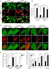
Overexpression of VDAC1 abrogates the mitochondrial protective function of miR-7. A and B, overexpression of VDAC1 abolishes the cytoprotective effect of miR-7 against MPP+-induced cell death. SH-SY5Y cells were co-transfected as indicated above. After 48 h, cells were treated with 2 mm MPP+ for 24 h followed by labeling of dead cells with PI. Representative images of MPP+-treated samples are shown in A. Arrows indicate dual (GFP and PI) positive cells that were counted as dead cells. Quantification of images is shown in B. C and D, overexpression of VDAC1 inhibits miR-7-induced decrease in mitochondrial calcium efflux. Plasmid pEGFP-C1 was also co-transfected in all samples, followed by treatment with 2 mm MPP+ for 12 h. Quantification of Rhod2-AM fluorescent intensities was performed from at least 10 cells among GFP-positive cells in each sample. These results (in A–D) are representatives of three separate experiments. E, overexpression of VDAC1 abolishes miR-7-induced decrease in intracellular ROS production. SH-SY5Y cells were transfected as indicated above. Forty-eight hours after transfection, cells were treated with the indicated concentrations of MPP+ for 12 h, and ROS levels were measured. This result (in E) is a representative of three separate experiments performed in triplicates for each group. Data are shown as the means ± S.E. ***, p < 0.005.

Similar articles

Cited by

References

    1. Abou-Sleiman P. M., Muqit M. M., and Wood N. W. (2006) Expanding insights of mitochondrial dysfunction in Parkinson's disease. Nat. Rev. Neurosci. 7, 207–219 - PubMed
    1. Duchen M. R. (2004) Mitochondria in health and disease: perspectives on a new mitochondrial biology. Mol. Aspects Med. 25, 365–451 - PubMed
    1. Exner N., Lutz A. K., Haass C., and Winklhofer K. F. (2012) Mitochondrial dysfunction in Parkinson's disease: molecular mechanisms and pathophysiological consequences. EMBO J. 31, 3038–3062 - PMC - PubMed
    1. Dias V., Junn E., and Mouradian M. M. (2013) The role of oxidative stress in Parkinson's disease. J. Parkinsons Dis. 3, 461–491 - PMC - PubMed
    1. Subramaniam S. R., and Chesselet M. F. (2013) Mitochondrial dysfunction and oxidative stress in Parkinson's disease. Prog. Neurobiol. 106–107, 17–32 - PMC - PubMed

Publication types

MeSH terms

Substances

LinkOut - more resources