Case Reports
. 2016 Jan 22:10:20.
doi: 10.1186/s13256-015-0783-5.

Relevance of frozen sections and serum markers in invasive squamous cell carcinoma arising from ovarian mature cystic teratoma: two case reports

Affiliations
Case Reports

Relevance of frozen sections and serum markers in invasive squamous cell carcinoma arising from ovarian mature cystic teratoma: two case reports

Yuki Tazo et al. J Med Case Rep. .

Abstract

Background: Ovarian mature cystic teratoma (MCT) is a common neoplasm in women. While malignant transformation of MCT is relatively rare, squamous cell carcinoma is the most frequent malignant neoplasm arising from MCT. Some tumor markers have been reported to be useful for prediction of MCT malignant transformation prior to operation. However, widely accepted use of these markers remains to be established. In the present study, we report the usefulness of frozen section assessment during operation, as well as preoperative measurement of tumor marker levels.

Case presentation: We present two cases of squamous cell carcinoma arising from ovarian MCT. The first case was a 45-year-old Asian woman referred to our hospital after her periodical company medical checkup, due to possible ovarian tumor. Image analysis suggested a dermoid cyst, and left salpingo-oophorectomy was performed. Because the cyst was histologically diagnosed as an invasive squamous cell carcinoma arising from an MCT, our patient underwent an additional preventative operation. The TNM classification and FIGO stage were T1aNXM0 and Ia, respectively. The second case was a 53 -year-old Asian woman who visited our hospital due to complaints of abdominal pain and urinary retention. Image analysis and laboratory data showing high serum levels of SCC antigen (normal range: < 1.5 ng/mL) and CA19-9 (normal range: < 37 U/mL), which strongly suggested malignant transformation of MCT. Frozen sections obtained during the operation were histologically analyzed to confirm malignancy, and our patient underwent an additional operation. The TNM classification and FIGO stage were T1aNXM0 and Ia, respectively.

Conclusions: We report the usefulness of frozen section assessment during operation, as well as preoperative measurement of tumor marker levels.


Figures
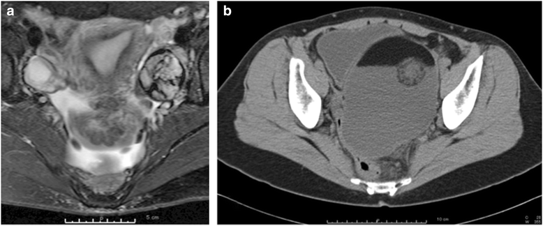
Fig. 1
Fig. 1
Image analysis. a Gadolinium-enhanced T1-weighted magnetic resonance imaging scan of the left ovary shows a cystic lesion, approximately three-fourths of which is occupied by an irregularly enhanced mass. b An enhanced computed tomography scan shows a large intra-abdominal cystic lesion occupying the pelvic cavity. Focal intracystic irregular enhancement is noted
Fig. 2
Fig. 2
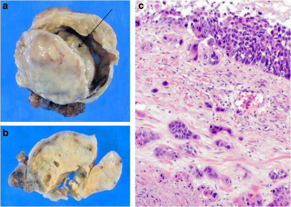
Gross findings and microscopic findings of case 1. a The inner surface of the cyst wall is irregularly thickened. A firm nodule is located within the cyst (arrow). b The cut surface of the cyst contains yellowish-gray, firm areas, and a few cystic cavities are embedded within. c A hematoxylin and eosin-stained section shows scattered islands of invasive squamous cell carcinoma (× 200)
Fig. 3
Fig. 3
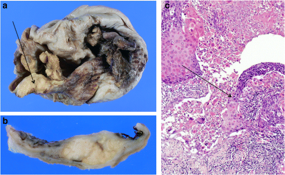
Gross findings and microscopic findings of case 2. a The wall of the cyst is thickened (arrow). b The cut surface of the cyst shows nodular protrusions. c A hematoxylin and eosin-stained section of the nodular area shows the boundary between the dysplastic keratinocytes lining the cyst internally, and the invasive squamous cell carcinoma (arrow) (× 100)

Similar articles

Cited by

References

    1. Iwasa A, Oda Y, Kaneki E, Ohishi Y, Kurihara S, Yamada T, et al. Squamous cell carcinoma arising in mature cystic teratoma of the ovary: an immunohistochemical analysis of its tumorigenesis. Histopathol. 2007;51:98–104. doi: 10.1111/j.1365-2559.2007.02727.x. - DOI - PubMed
    1. Hackethal A, Brueggmann D, Bohlmann MK, Franke FE, Tinneberg HR, Münstedt K. Squamous-cell carcinoma in mature cystic teratoma of the ovary: systematic review and analysis of published data. Lancet Oncol. 2008;9:1173–80. doi: 10.1016/S1470-2045(08)70306-1. - DOI - PubMed
    1. Tangjitgamol S, Manusirivithaya S, Sheanakul C, Leelahakorn S, Thawaramara T, Jesadapatarakul S. Squamous cell carcinoma arising from dermoid cyst: case reports and review of literature. Int J Gynecol Cancer. 2003;13:558–63. doi: 10.1046/j.1525-1438.2003.13312.x. - DOI - PubMed
    1. Dos Santos L, Mok E, Iasonos A, Park K, Soslow RA, Aghajanian C, et al. Squamous cell carcinoma arising in mature cystic teratoma of the ovary: a case series and review of the literature. Gynecol Oncol. 2007;105:321–4. doi: 10.1016/j.ygyno.2006.12.008. - DOI - PubMed
    1. Mori Y, Nishii H, Takabe K, Shinozaki H, Matsumoto N, Suzuki K, et al. Gynecol Oncol. 2003;90:338–341. doi: 10.1016/S0090-8258(03)00259-2. - DOI - PubMed

Publication types

Supplementary concepts