. 2016 Feb;73(2):68-82.
doi: 10.1002/cm.21275. Epub 2016 Feb 22.

Selective localization of myosin-I proteins in macropinosomes and actin waves

Affiliations

Selective localization of myosin-I proteins in macropinosomes and actin waves

Hanna Brzeska et al. Cytoskeleton (Hoboken). 2016 Feb.

Abstract

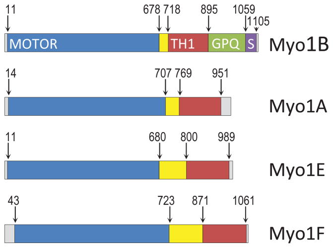
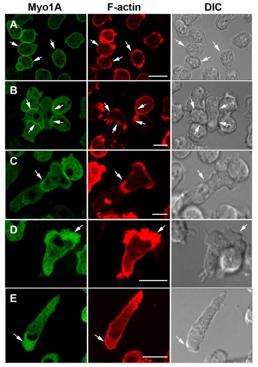
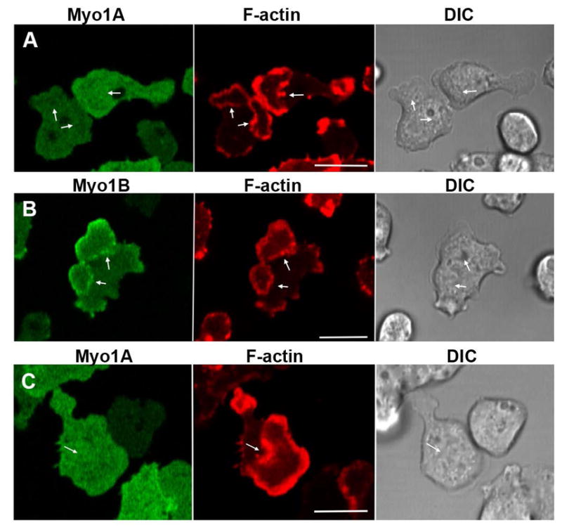
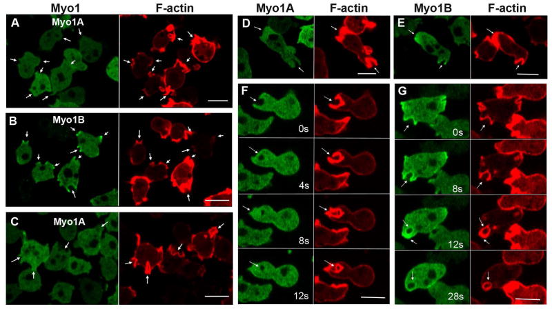
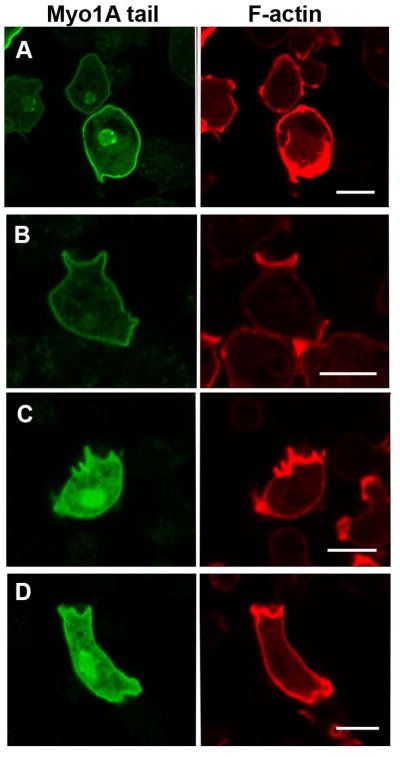
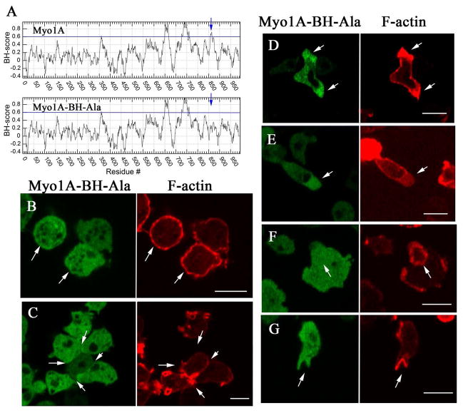
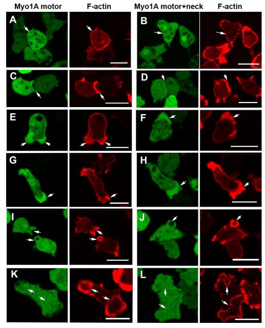
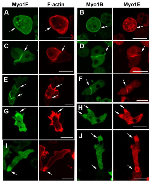
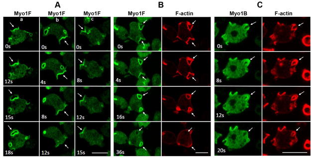
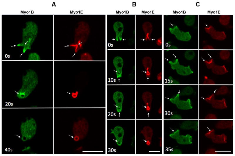
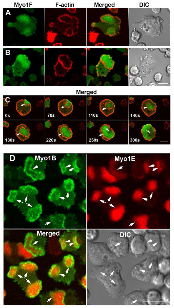
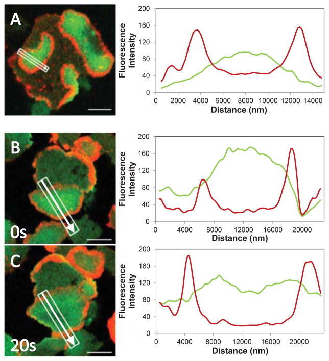
Class I myosins are widely expressed with roles in endocytosis and cell migration in a variety of cell types. Dictyostelium express multiple myosin Is, including three short-tailed (Myo1A, Myo1E, Myo1F) and three long-tailed (Myo1B, Myo1C, Myo1D). Here we report the molecular basis of the specific localizations of short-tailed Myo1A, Myo1E, and Myo1F compared to our previously determined localization of long-tailed Myo1B. Myo1A and Myo1B have common and unique localizations consistent with the various features of their tail region; specifically the BH sites in their tails are required for their association with the plasma membrane and heads are sufficient for relocalization to the front of polarized cells. Myo1A does not localize to actin waves and macropinocytic protrusions, in agreement with the absence of a tail region which is required for these localizations of Myo1B. However, in spite of the overall similarity of their domain structures, the cellular distributions of Myo1E and Myo1F are quite different from Myo1A. Myo1E and Myo1F, but not Myo1A, are associated with macropinocytic cups and actin waves. The localizations of Myo1E and Myo1F in macropinocytic structures and actin waves differ from the localization of Myo1B. Myo1B colocalizes with F-actin in the actin waves and at the tips of mature macropinocytic cups whereas Myo1E and Myo1F are in the interior of actin waves and along the entire surface of macropinocytic cups. Our results point to different mechanisms of targeting of short- and long-tailed myosin Is, and are consistent with these myosins having both shared and divergent cellular functions.

Keywords: BH site; actin waves; macropinocytosis; membrane binding; unconventional myosins.


Figures

Fig. 1
Fig. 1. Myosin Is used in this study
The boundaries of the motor (blue), TH1 (red), GPQ (green) and SH3 (purple) regions are marked according to Cymobase [Kollmar, 2006; Odronitz and Kollmar, 2006, 2007] and based on Pfam v.28. The region between the motor domain and TH1 domain contains the neck that contains light chain binding site(s). See text for exact boundaries of Myo1A mutants.
Fig. 2
Fig. 2. Localizations of Myo1A that are similar to the localization of Myo1B
GFP-Myo1A and the F-actin marker, RFP-lifeact, were coexpressed in Myo1A-null cells. Myo1A is present at: (A) the plasma membrane of non-motile cells, (B) the sites of cell-cell contacts, (C) regions of cell-cell contact between chemotaxing cells, (D) in the cortex of pseudopods of randomly moving cells, and (E) at the front of chemotaxing cells. All the above localizations are similar to that for Myo1B. Panel A shows fixed cells, all other panels show live cells. Arrows point to the structures of interest. Bars are 10 μm.
Fig. 3
Fig. 3. Myo1A is absent from actin waves
GFP-Myo1A or GFP-Myo1B was coexpressed with RFP-lifeact in Myo1A-null or Myo1B-null cells. Actin waves were induced by 1 μM latrunculin. (A) Myo1A does not localize to actin waves in Myo1A-null cells. (B) Myo1B does localize to actin waves in Myo1A-null cells. (C) Myo1A does not localize to actin waves in Myo1B-null cells. Images of live cells are shown. Arrows indicate positions of actin waves identified by RFP-lifeact fluorescence. Bars are 10 μm.
Fig. 4
Fig. 4. Myo1A is absent from macropinocytic protrusions
GFP-Myo1A or GFP-Myo1B was co-expressed with RFP-lifeact in Myo1A-null and Myo1B-null cells, and their distributions observed in live cells undergoing macropinocytosis. (A) Myo1A does not localize to macropinocytic protrusions in Myo1A-null cells. (B) Myo1B does localize to macropinocytic protrusions in Myo1A-cells. (C) Myo1A does not localize to macropinocytic protrusions in Myo1B-null cells. (D) Myo1A does localize to a pseudopods but not to macropinocytic cups in Myo1A-null cells. (E) Myo1B localizes to pseudopod and to a macropinocytic cup in a Myo1A-null cell. (F) Formation of a macropinocytic cup in a Myo1B-null cell coexpressing Myo1A and lifeact. Images were taken at times indicated in the figure. Myo1A does not localize to the cup. (G) Formation of a macropinocytic cup in a Myo1B-null cell coexpressing Myo1B and lifeact. Myo1B does localize to the cup. Images were taken at times indicated in the figure. Arrows point to macropinocytic protrusions. Bars are 10 μm.
Fig. 5
Fig. 5. The Myo1A tail localizes to the plasma membrane
GFP-Myo1A tail and RFP-lifeact were coexpressed in Myo1A-null and Myo1B-null cells. (A) Myo1A tail in non-motile Myo1A-null cells. (B) Myo1A tail in a non-motile Myo1B-null cell. (C) Myo1A tail in a randomly moving Myo1B-null cell. (D) Myo1A tail in a chemotaxing Myo1B-null cell. (A) and (B) show fixed cells. (C) and (D) show live cells. Bars are 10 μm.
Fig. 6
Fig. 6. Plasma membrane localization of Myo1A requires the BH site
GFP-Myo1A-BH-Ala and RFP-lifeact were coexpressed in Myo1A-null cells. (A) BH plots of Myo1A and Myo1A-BH-Ala mutant run with window 19. The position of the BH peak is marked with an arrow. The regions with BH values above the horizontal line at 0.6 are considered to be positive BH peaks and the mutated BH site in the tail is marked with an arrow. (B) Myo1A-BH-Ala does not localize to the plasma membrane of non-motile cells. (C) Myo1A-BH-Ala does not localize to the sites of cell-cell contacts of non-motile cells. (D) Myo1A-BH-Ala localizes to pseudopods. (E) Myo1A-BH-Ala localizes to the front of chemotaxing cells. (F) Myo1A-BH-Ala does not localize to actin waves. (G) Myo1A-BH-Ala does not localize to macropinocytic cups. Arrows point to the structures of interest. (B) shows fixed cells, and (C)–(G) show live cells. Bars are 10 μm.
Fig. 7
Fig. 7. Myo1A motor domain localizes to pseudopods and the cell front
Myo1A GFP-Motor or GFP-Motor+Neck was coexpressed with RFP-lifeact in GFP-A-null cells. Both Myo1A truncation mutants are absent from the plasma membrane of non-motile cells (A, B) and sites of cell-cell contacts (C, D). Both Myo1A truncation mutants localize to pseudopods (E, F) and to the front of chemotaxing cells (G, H). Like full length Myo1A, neither Myo1A mutant localizes to either macropinocytic cups (I, J) or to actin waves (K, L). (A) and (B) show non-motile fixed cells, the other panels show live cells. Arrows point to sites of interest. Bars are 10 μm.
Fig. 8
Fig. 8. Localizations of Myo1F and Myo1E that are similar to the localization of Myo1A
GFP-Myo1F and RFP-Myo1E were coexpressed with RFP-lifeact or RFP-Myo1E, respectively, in Myo1B-null cells. Myo1F and Myo1E, as well as Myo1B, localize to: (A, B) the plasma membrane, (C, D) sites of cell-cell contacts, (E, F) sites of cell-cell contact between chemotaxing cells, (G, H) pseudopods, and (I, J) the front of chemotaxing cells. (A and B) show fixed non-motile cells, and (C–J) show live cells. Arrows point to the sites of interest. Bars are 10 μm.
Fig. 9
Fig. 9. Localization of Myo1F in macropinocytic structures
GFP-Myo1F or GFP-Myo1B were coexpressed with RFP-lifeact in Myo1B-null cells. Images of live cells were taken at the times indicated in the panels. (A) Localization of Myo1F during formation of various kinds of extensions. Column (a) flat protrusion turning into a macropinocytic cup. Column (b) flat protrusion disappearing without turning into a macropinocytic cup. Column (c) macropinocytic cup turning into a flat protrusion. (B) Localization of Myo1F and F-actin during internalization of a pinocytic vesicle. (C) Localization of Myo1B and F-actin during internalization of a pinocytic vesicle. Arrows point to the locations of interest (see text). Bars are 10 μm.
Fig. 10
Fig. 10. Different Localizations of Myo1E and Myo1B during macropinocytosis
RFP-Myo1E and GFP-Myo1B were coexpressed in Myo1B-null cells. Images of live cells were taken at times indicated in the panels. Different characterictics of macropinocytic structures are shown in panels (A), (B) and (C), see text for description. Arrows point to the structures of interest. Bars are 10 μm.
Fig. 11
Fig. 11. Myo1F and Myo1E localize to the interior of actin waves
Cells were cotransfected with GFP-Myo1F and RFP-lifeact (A–C) or with RFP-Myo1E and GFP-Myo1B (D). Actin waves were induced with 1 μM latrunculin. (A) Myo1F-null cells expressing Myo1F and lifeact. (B) Myo1B-null cells expressing Myo1F and lifeact. (C) AX2 cells expressing Myo1F and lifeact, snapshots of wave propagation taken at times indicated in the panel. (D) Myo1B-null cells expressing Myo1E and Myo1B. Images of live cells are shown. Arrows point to actin waves. Bars are 10 μm.
Fig. 12
Fig. 12. Linescans of Myo1F and F-actin fluorescence in actin waves
GFP-Myo1F and RFP-lifeact were coexpressed in Myo1B-null cells and actin waves were induced with 1 μM latrunculin. Merged cell images are shown at the left and line scans of GFP-Myo1F and RFP-lifeact fluorescence are shown at the right. (A) Cell with multiple waves, the Myo1F fluorescence peak is located in the middle of the region encircled by a wave. This image corresponds to supplemental movie M2. (B) and (C) actin wave in the same cell at 0 s and 20 s. During wave expansion Myo1F fluorescence decayed in the middle of the wave. The white rectangle shows the region scanned, the arrow indicates the direction of scanning. Images of live cells are shown. Bars are 10 μm.

Similar articles

Cited by

References

    1. Bagorda A, Mihaylov VA, Parent CA. Chemotaxis: moving forward and holding on to the past. Thromb Haemost. 2006;95:12–21. - PubMed
    1. Benesh AE, Nambiar R, McConnell RE, Mao S, Tabb DL, Tyska MJ. Differential localization and dynamics of class I myosins in the enterocyte microvillus. Mol Biol Cell. 2010;21:970–978. - PMC - PubMed
    1. Berg JS, Powell BC, Cheney RE. A millennial myosin census. Mol Biol Cell. 2001;12:780–794. - PMC - PubMed
    1. Bloomfield G, Traynor D, Sander SP, Veltman DM, Pachebat JA, Kay RR. Neurofibromin controls macropinocytosis and phagocytosis in Dictyostelium. eLife. 2015;4 - PMC - PubMed
    1. Bond LM, Brandstaetter H, Kendrick-Jones J, Buss F. Functional roles for myosin 1c in cellular signaling pathways. Cell Signal. 2013;25:229–235. - PMC - PubMed

Publication types

LinkOut - more resources