. 2016 Jan 22:23:14.
doi: 10.1186/s12929-016-0234-7.

Surface vimentin is critical for the cell entry of SARS-CoV

Affiliations

Surface vimentin is critical for the cell entry of SARS-CoV

Yvonne Ting-Chun Yu et al. J Biomed Sci. .

Abstract

Background: Severe acute respiratory syndrome coronavirus (SARS-CoV) caused a global panic due to its high morbidity and mortality during 2002 and 2003. Soon after the deadly disease outbreak, the angiotensin-converting enzyme 2 (ACE2) was identified as a functional cellular receptor in vitro and in vivo for SARS-CoV spike protein. However, ACE2 solely is not sufficient to allow host cells to become susceptible to SARS-CoV infection, and other host factors may be involved in SARS-CoV spike protein-ACE2 complex.

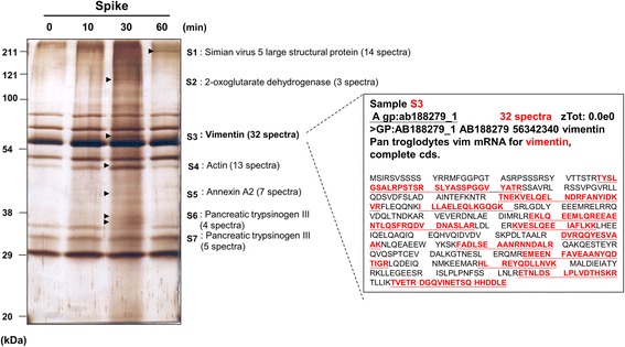
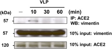
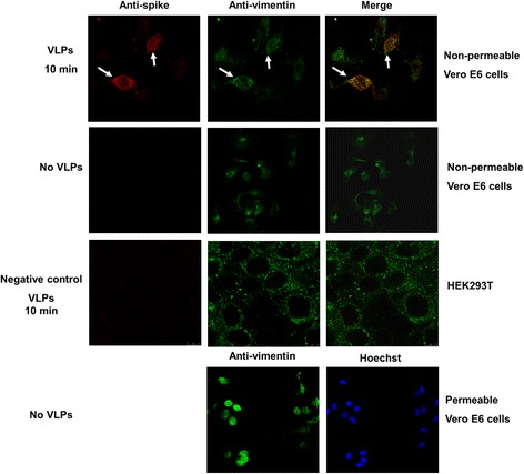
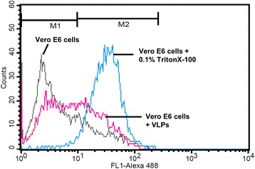
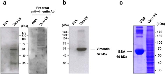
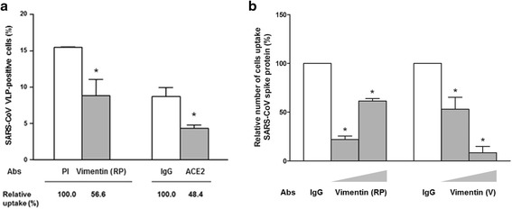
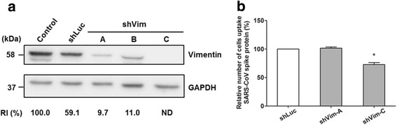
Results: A host intracellular filamentous cytoskeletal protein vimentin was identified by immunoprecipitation and LC-MS/MS analysis following chemical cross-linking on Vero E6 cells that were pre-incubated with the SARS-CoV spike protein. Moreover, flow cytometry data demonstrated an increase of the cell surface vimentin level by 16.5 % after SARS-CoV permissive Vero E6 cells were treated with SARS-CoV virus-like particles (VLPs). A direct interaction between SARS-CoV spike protein and host surface vimentin was further confirmed by far-Western blotting. In addition, antibody neutralization assay and shRNA knockdown experiments indicated a vital role of vimentin in cell binding and uptake of SARS-CoV VLPs and the viral spike protein.

Conclusions: A direct interaction between vimentin and SARS-CoV spike protein during viral entry was observed. Vimentin is a putative anti-viral drug target for preventing/reducing the susceptibility to SARS-CoV infection.


Figures

Fig. 1
Fig. 1
Vimentin as a cross-linked protein in association with the SARS-CoV spike protein-ACE2 complex. Vero E6 cells at 8 × 106 were pre-incubated with SARS-CoV spike protein (1 μg/106 cells) at 4 °C for various time periods as indicated followed by a treatment with the thiol-cleavable cross-linker DTSSP. Cell lysates were harvested and subjected to immunoprecipitation with anti-ACE2 antibodies. Vero E6 cells being treated by SARS-CoV VLPs for 0 min was used as the negative control. The immunoprecipitates were incubated with protein sample buffer with the addition of 5 % β-mercaptoethenol and subjected to SDS-PAGE. Proteins separated on the polyacrylamide gel were visualized by silver staining. The protein bands marked S1 to S7 were collected for LC-MS/MS analysis and results were shown. The number of matched spectra of the protein fragments compared to the published data was stated in parentheses. The detailed analysis of S3 was shown in the inset, where sequence comparison with data banks indicated the S3 protein to be vimentin. Underlines show the peptide sequences of 32 spectra
Fig. 2
Fig. 2
Vimentin involved in the SARS-CoV spike-ACE2 complex on plasma membrane at different time points. Vero E6 cells were incubated with SARS-CoV VLPs for 10, 30 and 60 min at 37 °C. Cell lysates were then prepared and subjected to immunoprecipitation (IP) with anti-ACE2 antibodies, followed by Western blot (WB) analysis with anti-vimentin antibodies (Sigma). Cell lysates without the process of immunoprecipitation (10 % input) were analyzed in parallel with anti-vimentin and anti-ACE2 antibodies as the protein loading controls
Fig. 3
Fig. 3
Co-localization of the spike protein with vimentin in Vero E6 cells pre-treated with SARS-CoV VLPs. Vero E6 cells pre-treated with SARS-CoV VLPs for 10 min were fixed for immunofluorescence assay with anti-spike antibodies followed by AlexaFluor 594-conjugated goat anti-rabbit IgG and anti-vimentin antibodies followed by AlexaFluor 488-conjugated goat anti-mouse IgG antibodies as shown in red and green colors, respectively. Vero E6 cells without the pretreatment with SARS-CoV VLPs and ACE2-negative HEK293T cells treated with SARS-CoV VLPs for 10 min were used as controls. Hoechst staining was performed in parallel to localize cell nuclei in the field and the distribution of vimentin in cytosol was examined using permeable Vero E6 cells
Fig. 4
Fig. 4
An increased level of cell surface vimentin induced by SARS-CoV VLPs. Vero E6 cells suspended in FACS buffer were incubated with SARS-CoV VLPs for 10 min at 4 °C. The cells were then fixed and analyzed with anti-vimentin antibodies and AlexaFluor 488-conjugated secondary antibodies. The fluorescence was quantified by using a LSR-II Digital Flow Cytometer and the data were analyzed by using FlowJo software. Vero E6 cells without the pre-treatment of SARS-CoV VLPs were used as a negative control (M1 region) and Vero E6 cells permeabilized with 0.1 % TritonX-100 for 5 min were used as a positive control (M2 region)
Fig. 5
Fig. 5
Direct interaction between vimentin and SARS-CoV spike protein analyzed by far-Western blot analysis. a Far-Western blot analysis. Protein lysates prepared from Vero E6 cells were blotted on PVDF membrane and subjected to far-Western blot analysis with anti-V5 epitope antibodies (left panel) following a pre-incubation of the membrane with the purified V5-tagged recombinant SARS-CoV spike protein. The right panel showed the Vero E6 cells pre-incubated with anti-vimentin antibodies (Sigma) prior to the treatment of SARS-CoV spike protein. b Western blot analysis. The same set of protein lysates blotted on PVDF membrane was subjected to Western blot analysis with anti-vimentin antibodies to serve as a positive control. c Coomassie blue staining. Coomassie blue staining showed the total proteins on the gel. BSA loaded in parallel was used as a negative control
Fig. 6
Fig. 6
Anti-vimentin antibodies specifically diminished the uptake of SARS-CoV VLPs and spike protein by Vero E6 cells. a The effect of anti-vimentin antibodies and anti-ACE2 antibodies on the uptake of SARS-CoV VLPs. Vero E6 cells were pretreated with rabbit anti-vimentin antibodies RP (RP4002, Immuno Bioscience Corp.) or goat anti-ACE2 antibodies for 30 min prior to the 2-h incubation with SARS-CoV VLPs. The cells were then subjected to flow cytometry analysis for quantitation of the SARS-CoV VLP-positive cells as described in Methods. Rabbit pre-immune serum (PI), and goat IgG as indicated were used as antibody isotype controls in the neutralization experiments. The number of cells pretreated with isotype antibodies was normalized to 100 %, subsequently the relative uptake of SARS-CoV VLPs after antibody neutralization was calculated and shown underneath the bar chart. b The effect of anti-vimentin antibodies on the uptake of spike protein. Vero E6 cells were pretreated with increasing doses of rabbit anti-vimentin antibodies RP (RP4002, Immuno Bioscience Corp.) or mouse anti-vimentin antibodies V (V5255, Sigma) as indicated for 30 min prior to the 2-h incubation with SARS-CoV spike protein. SARS-CoV spike-positive cells were then quantitated by flow cytometry analysis. The number of cells uptake SARS-CoV spike protein after antibody neutralization was calculated and normalized against the number of cells pretreated with IgG control in each set of experiment. * indicates the P value < 0.05
Fig. 7
Fig. 7
Knockdown of vimentin expression to an undetectable level specifically reduced the uptake of SARS-CoV spike protein by Vero E6 cells. a Lentivirus-mediated knockdown of vimentin. Vero E6 cells infected by lentiviruses carrying shRNAs specific to vimentin (shVim-A, B, and C) were harvested at 4 days postinfection for Western blot analysis with antibodies against vimentin (Sigma). Cells without infection (Control) and cells infected by lentiviruses carrying shRNA against luciferase (shLuc) served as controls. GAPDH was used as an internal control. RI: relative intensity; ND: not detectable. b The effect of vimentin knockdown on the uptake of SARS-CoV spike protein. Vero E6 cells were infected by lentiviruses carrying shRNAs as indicated for 4 days prior to the 2-h incubation with SARS-CoV spike proteins. The cells were then subjected to flow cytometry analysis for quantitation of the SARS-CoV spike-positive cells. The number of cells uptake SARS-CoV spike proteins after vimentin knockdown was calculated and normalized to the number of cells pretreated with lentiviruses carrying shRNA against luciferase (shLuc). * indicates the P value < 0.05
Fig. 8
Fig. 8
Simplified diagram of the involvement of vimentin within SARS-CoV spike-ACE2 complex during cell entry. The dashed circle indicates the spike-ACE2 complex on the cellular membrane, and the blue dotted arrow demonstrates the possible pathway when vimentin translocates from cytosol to membrane

Similar articles

Cited by

References

    1. Peiris JSM, Lai ST, Poon LLM, Guan Y, Yam LYC, Lim W, et al. Coronavirus as a possible cause of severe acute respiratory syndrome. Lancet. 2003;361:1319–25. doi: 10.1016/S0140-6736(03)13077-2. - DOI - PMC - PubMed
    1. Groneberg DA, Zhang L, Welte T, Zabel P, Chung KF. Severe acute respiratory syndrome: global initiatives for disease diagnosis. Q J Med. 2003;96:845–52. doi: 10.1093/qjmed/hcg146. - DOI - PMC - PubMed
    1. Ksiazek TG, Erdman D, Goldsmith CS, Zaki SR, Peret T, Emery S, et al. A novel coronavirus associated with severe acute respiratory syndrome. N Engl J Med. 2003;348:1953–66. doi: 10.1056/NEJMoa030781. - DOI - PubMed
    1. Drosten C, Günther S, Preiser W, van der Werf S, Brodt HR, Becker S, et al. Identification of a novel coronavirus in patients with severe acute respiratory syndrome. N Engl J Med. 2003;348:1967–76. doi: 10.1056/NEJMoa030747. - DOI - PubMed
    1. Li W, Moore MJ, Vasilieva N, Sui J, Wong SK, Berne MA, et al. Angiotensin-converting enzyme 2 is a functional receptor for the SARS coronavirus. Nature. 2003;426:450–4. doi: 10.1038/nature02145. - DOI - PMC - PubMed

Publication types

MeSH terms