. 2016 Mar;117(3):401-19.
doi: 10.1093/aob/mcv187. Epub 2016 Jan 22.

Cell wall matrix polysaccharide distribution and cortical microtubule organization: two factors controlling mesophyll cell morphogenesis in land plants

Affiliations

Cell wall matrix polysaccharide distribution and cortical microtubule organization: two factors controlling mesophyll cell morphogenesis in land plants

P Sotiriou et al. Ann Bot. 2016 Mar.

Abstract

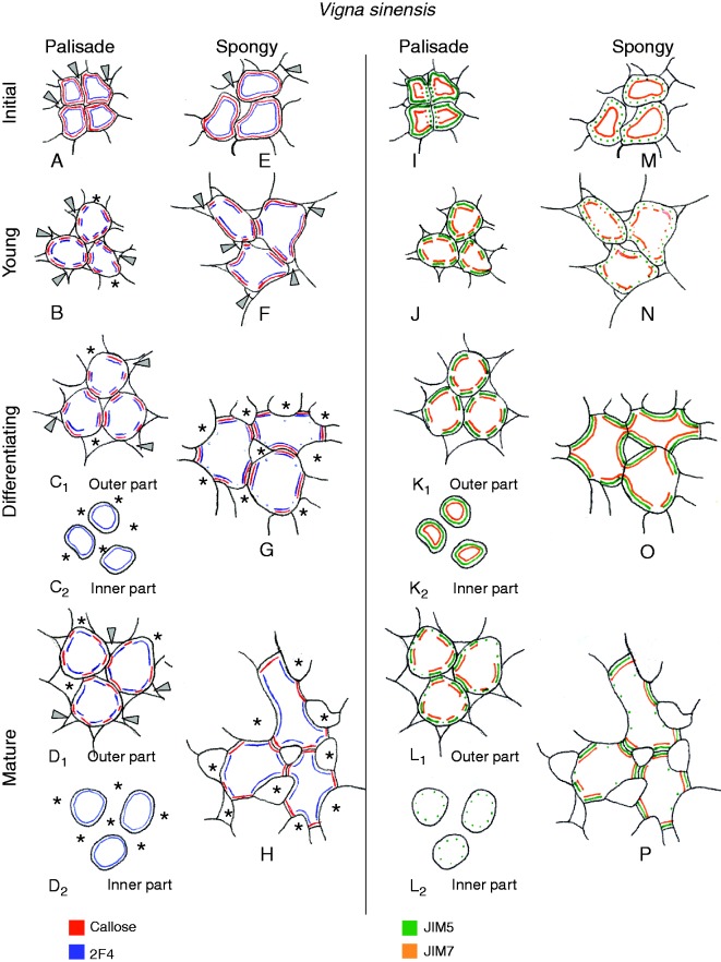
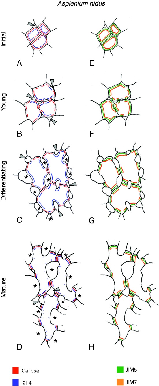
Background and aims: This work investigates the involvement of local differentiation of cell wall matrix polysaccharides and the role of microtubules in the morphogenesis of mesophyll cells (MCs) of three types (lobed, branched and palisade) in the dicotyledon Vigna sinensis and the fern Asplenium nidus.

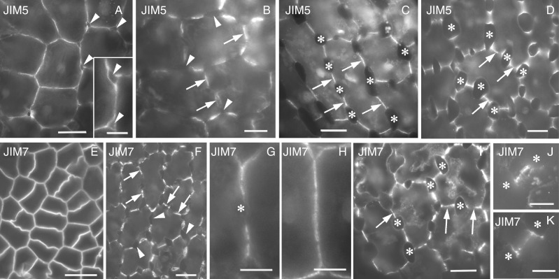
Methods: Homogalacturonan (HGA) epitopes recognized by the 2F4, JIM5 and JIM7 antibodies and callose were immunolocalized in hand-made leaf sections. Callose was also stained with aniline blue. We studied microtubule organization by tubulin immunofluorescence and transmission electron microscopy.

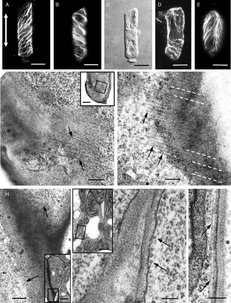
Results: In both plants, the matrix cell wall polysaccharide distribution underwent definite changes during MC differentiation. Callose constantly defined the sites of MC contacts. The 2F4 HGA epitope in V. sinensis first appeared in MC contacts but gradually moved towards the cell wall regions facing the intercellular spaces, while in A. nidus it was initially localized at the cell walls delimiting the intercellular spaces, but finally shifted to MC contacts. In V. sinensis, the JIM5 and JIM7 HGA epitopes initially marked the cell walls delimiting the intercellular spaces and gradually shifted in MC contacts, while in A. nidus they constantly enriched MC contacts. In all MC types examined, the cortical microtubules played a crucial role in their morphogenesis. In particular, in palisade MCs, cortical microtubule helices, by controlling cellulose microfibril orientation, forced these MCs to acquire a truncated cone-like shape. Unexpectedly in V. sinensis, the differentiation of colchicine-affected MCs deviated completely, since they developed a cell wall ingrowth labyrinth, becoming transfer-like cells.

Conclusions: The results of this work and previous studies on Zea mays (Giannoutsou et al., Annals of Botany 2013; 112: : 1067-1081) revealed highly controlled local cell wall matrix differentiation in MCs of species belonging to different plant groups. This, in coordination with microtubule-dependent cellulose microfibril alignment, spatially controlled cell wall expansion, allowing MCs to acquire their particular shape.

Keywords: 2F4; Asplenium nidus; Callose; JIM5; JIM7; Vigna sinensis; cell contacts; cell wall; homogalacturonan.; microtubules; morphogenesis of photosynthetic cells; pectin epitopes.


Figures

F<sc>ig</sc>. 1.
Fig. 1.
MCs of V. sinensis as they appear under the light microscope after staining with toluidine blue. (Α) Transverse section of a young leaf in which the palisade MC initials (upper part of the mesophyll) and the spongy MC initials (lower part of the mesophyll) can be observed. (B, C) Transverse (B) and paradermal (C) sections of palisade MC initials. The arrowheads in (B, C), as well as those in (G), point to intercellular spaces. (D) Spongy MC initials in a paradermal section. Asterisks mark intercellular spaces. (E, F) Median transverse sections of differentiating palisade MCs. The asterisks in (E–J) mark intercellular spaces. (G, H) Paradermal sections passing through the outermost (G) and innermost (H) parts of the differentiating palisade MCs. (I, J) Differentiating (I) and mature (J) spongy MCs in median paradermal sections. EC, epidermal cell. Scale bars: (A, I, J) = 20 μm; (B, C, D, G, H) = 10 μm; (E, F) = 5 μm.
F<sc>ig</sc>. 2.
Fig. 2.
MC contacts of V. sinensis as seen by TEM. (A) An entire contact between mature MCs. Note the absence of plasmodesmata. Asterisks mark intercellular spaces. (B) Part of a contact between differentiating MCs traversed by plasmodesmata (arrowheads), where the process of occlusion has not yet been concluded. The arrows point to endoplasmic reticulum elements. (C) Part of an MC contact in which a local cell wall thickening has formed (arrowhead), probably in the region of occluded plasmodesmata. Similar cell wall thickenings are shown by the arrowheads in (A). The arrow in (C) shows endoplasmic reticulum adjacent to the cell wall thickening. Scale bars = 1 μm.
F<sc>ig</sc>. 3.
Fig. 3.
Young palisade MCs as seen after tubulin immunolabelling (A, B, D, E), differential interference contrast (DIC) optics (C) and TEM (F–J). The double arrow in (A) defines the longitudinal axis of the MCs, which are oriented with their outermost part towards the top of the figure. (A, B) Optical sections passing through the two opposite anticlinal faces of the same MC. The helical organization of the cortical microtubules is obvious. (C) The cell shown in (A) and (B) in DIC optics. (D) Young MC as seen by CLSM after tubulin immunolabelling. Note the different orientation of the microtubules in the outermost and innermosts part of the cell. Maximum projection of an image stack of ten optical sections taken by CLSM. (E) Organization of cortical microtubules in the upper part of the anticlinal faces of a young palisade MC. (F) Higher magnification of the region delimited by the inset frame. The arrows point to a cortical microtubule bundle oriented almost perpendicularly to the cell’s long axis. (F inset) Innermost part of a young palisade MC. (G) Bundle of cortical microtubules (arrows) in a young palisade MC. The cellulose microfibrils (white dashed lines) are co aligned with the microtubules. (H) Region outlined by the frame in the inset at higher magnification. The arrows point to cortical microtubule bundles. Note their different orientations in the two adjacent cells. (H inset) Section passing through the innermost part of two adjacent young palisade MCs. (I) Region demarcated by the frame in the inset at higher magnification. The arrows point to a bundle of cortical microtubules oriented perpendicularly to the MC axis. (I inset) Young palisade MC in a longitudinal section. (J) Cortical cytoplasmic region of a young palisade MC. Arrows indicate a bundle of cortical microtubules oriented parallel to the cell’s long axis. The asterisk marks an intercellular space. Scale bars: (A–E) = 5 μm; (F–J) = 200 nm; (F inset) = 3 μm; (H, I inset) = 2 μm.
F<sc>ig</sc>. 4.
Fig. 4.
Colchicine-treated MCs of V. sinensis as they appear in the light microscope after toluidine blue staining (A, B), by TEM (C, D, F–I) and after tubulin immunolabelling (E). The cells were treated with colchicine at 0·08 % (w/v) for 48–96 h. (A, B) Treated MCs in transverse (A) and paradermal (B) sections. The complete deviation of cell morphogenesis and the absence of well-developed intercellular spaces in the mesophyll are obvious. Arrowheads in (A) show aberrant sieve elements (Galatis, 1991). EC, epidermal cell. (C) Treated palisade MC in a longitudinal section (in its anticlinal cell walls, indicated by arrows, numerous local cell wall ingrowths can be seen). P, plastid. (D) Anticlinal cell wall region of (C) at higher magnification. Arrowheads point to electron-translucent material at the borders of the cell wall ingrowths. (E) Network of atypical tubulin polymers in the cortical cytoplasm of a treated MC. (F–I) Tubulin/colchicine paracrystals (arrows) in transverse (F) and longitudinal (G–I) sections. Arrowheads in (I) mark endoplasmic reticulum elements. Scale bars: (A) = 20 μm; (B) = 10 μm; (C, E) = 5 μm; (D) = 500 nm; (F–I) = 200 nm.
F<sc>ig</sc>. 5.
Fig. 5.
Callose detection after staining with aniline blue (A–E) and immunolabelling of the 2F4 HGA epitope (F–J) in paradermal optical sections of palisade MCs of V. sinensis. Arrows point to MC contacts and arrowheads or asterisks indicate intercellular spaces. (A) MC initials. (B, C) Optical sections passing through the median plane (B) and the inner periclinal cell wall (C) of a group of young MCs. (D, E) Optical sections through the outermost part of differentiating (D) and mature (E) palisade MCs. (F) MC initials. (G) Young MCs in a median optical plane. (H) Optical section passing through the innermost part of differentiating palisade MCs. (I, J) Mature MCs in an optical plane passing through their outermost (I) and innermost (J) parts. Scale bars = 10 μm.
F<sc>ig</sc>. 6.
Fig. 6.
Distribution of HGA epitopes recognized by JIM5 (A–F) and JIM7 (G–I) antibodies in paradermal optical sections of palisade MCs of V. sinensis. Arrows point to MC contacts and arrowheads and asterisks indicate intercellular spaces. (A) MC initials displaying intense JIM5 fluorescent signal at parental cell walls, and very weak signal at young daughter cell walls (open arrows). (B) Area of the palisade parenchyma where groups of MC initials are delimited by an intense JIM5 fluorescent signal. (C) Very young MCs in median optical section. (D, E) Optical sections through the outermost (D) and innermost (E) parts of differentiating MCs. (F) Optical section passing through the outermost part of mature MCs. (G–I) Young (G), differentiating (H) and mature (I) MCs. Open arrows in G point to daughter cell walls. Scale bars = 10 μm.
F<sc>ig</sc>. 7.
Fig. 7.
Callose localization after aniline blue staining (A–C) and immunolocalization of HGA epitopes recognized by 2F4 (D–F), JIM5 (G–I) and JIM7 (J–L) antibodies in paradermal sections of spongy MCs of V. sinensis. Arrows point to MC contacts and arrowheads and asterisks indicate intercellular spaces. (D) MC initials; (A, G, J) young MCs; (B, E, H, K); differentiating MCs; (C, F, I, L) mature MCs. Scale bars = 10 μm.
F<sc>ig</sc>. 8.
Fig. 8.
Diagrammatic representation of distributions of callose and HGA epitopes recognized by 2F4, JIM5 and JIM7 antibodies throughout cell walls of palisade and spongy MCs of V. sinensis at successive stages of differentiation. The drawings represent exact copies of MC cell walls in paradermal sections. Arrowheads and asterisks indicate intercellular spaces. Cells in (A, B, E, F) and (I, J, M, N) are shown at a higher magnification that those in (C, D, G, H) and (K, L, O, P).
F<sc>ig</sc>. 9.
Fig. 9.
MCs of A. nidus as seen by differential interference contrast (DIC) optics (A, D) after immunolabelling of tubulin (B, C, E) and by TEM (F–H). (A) Mature MCs. Asterisks show intercellular spaces. (B–D) Organization of cortical microtubules as shown in optical sections passing through the surface (B) and the middle (C) of a young MC. They form bundles at the regions of the newly formed cellular constrictions. The latter can be observed in (D). (E) Microtubule immunolabelling in a group of differentiating MCs. Tubulin immunofluorescent signal is located only in regions of newly formed cellular constrictions. Asterisks and arrowheads show newly formed intercellular spaces. (F) MC contact as seen by TEM. Note the absence of plasmodesmata. Asterisks mark intercellular spaces. (G) The arrow points to an area of an MC contact that may be a primary pit field. Notice the lack of plasmodesmata. (H) Part of an MC contact. Arrowheads point to plasmodesmata that have been blocked by electron-dense material. Scale bars: (A) = 20 μm; (B–E) = 10 μm; (F–H) = 1 μm.
F<sc>ig</sc>. 10.
Fig. 10.
Callose localization after staining with aniline blue (A–C) and immunolabelling of 2F4 HGA epitope (D–F) in young (A, D), differentiating (B, E) and mature (C, F) MCs of A. nidus. Arrows show MC contacts; the arrowhead in (C) and asterisks show intercellular spaces. In (F) arrows 1 point to optical sections passing through the middle of cell contacts, while arrows 2 point to optical sections passing through the margins of cell contacts. Scale bars = 20 μm.
F<sc>ig</sc>. 11.
Fig. 11.
Immunolocalization of HGA epitopes recognized by JIM5 (A–D) and JIM7 (E–K) antibodies in MCs of A. nidus. (A) MC initials. Part of the cell wall is shown at higher magnification in the inset. Arrowheads point to intercellular spaces. (B–D) Young (B), differentiating (C) and mature (D) MCs. Arrows indicate MC contacts and arrowheads and asterisks indicate intercellular spaces. (E) MC initials. (F) Young MCs. Arrows point to MC contacts and arrowheads to intercellular spaces. (G, H) Cell wall of a young MC in external (G) and median (H) optical section. The asterisk in (G) marks the region of a newly formed intercellular space. In (H) the intercellular space formation has not yet progressed. (I) Differentiating MCs. Arrows point to MC contacts and asterisks indicate intercellular spaces. (J, K) Cell contact of a mature MC in external (J) and median (K) optical section. Asterisks mark intercellular spaces. Scale bars: (A, B, E, F, G, H, J, K) = 10 μm; (C, D, I) = 20 μm; (A inset) = 5 μm.
F<sc>ig</sc>. 12.
Fig. 12.
Diagrammatic representation illustrating the distribution of callose and of HGA epitopes recognized by 2F4, JIM5 and JIM7 antibodies in the cell walls of MCs of A. nidus at successive stages of differentiation. Arrowheads and asterisks indicate intercellular spaces. The drawings represent exact copies of MC cell walls in paradermal sections. Cells in (A, B, E, F) are shown at a higher magnification that those in (C, D, G, H).

Similar articles

Cited by

References

    1. Altartouri Β, Geitmann Α. 2014. Understanding plant cell morphogenesis requires real-time monitoring of cell wall polymers. Current Opinion in Plant Biology 23C: 76–82. - PubMed
    1. Apostolakos P, Galatis B, Katsaros C, Schnepf E. 1990. Tubulin conformation in microtubule-free cells of Vigna sinensis: an immunofluorescent and electron microscope study. Protoplasma 154: 132–143.
    1. Apostolakos P, Galatis B, Panteris E. 1991. Microtubules in cell morphogenesis and intercellular space formation in Zea mays leaf mesophyll and Pilea cadierei epithem. Journal of Plant Physiology 137: 591–601.
    1. Bacic A, Fincher GB, Stone BA. 2009. Chemistry, biochemistry, and biology of (1→3)-β-glucans and related polysaccharides. Amsterdam: Academic Press.
    1. Baluška F, Liners F, Hlavačka A, et al. 2005. Cell wall pectins and xyloglucans are internalized into dividing root cells and accumulate within cell plates during cytokinesis. Protoplasma 225: 141–155. - PubMed

MeSH terms