. 2016 Mar:490:17-26.
doi: 10.1016/j.virol.2016.01.002. Epub 2016 Jan 21.

Modulating immunogenic properties of HIV-1 gp41 membrane-proximal external region by destabilizing six-helix bundle structure

Affiliations

Modulating immunogenic properties of HIV-1 gp41 membrane-proximal external region by destabilizing six-helix bundle structure

Saikat Banerjee et al. Virology. 2016 Mar.

Abstract

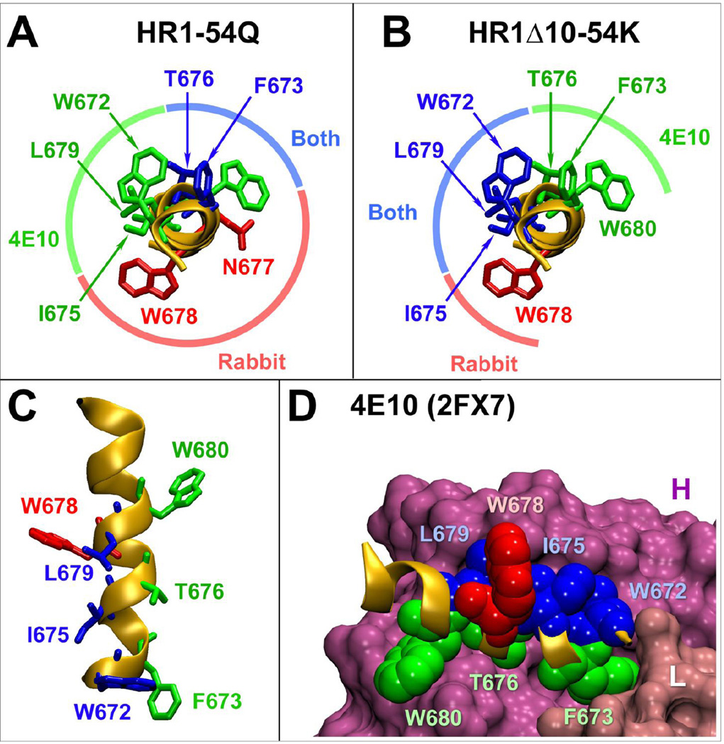
The C-terminal alpha-helix of gp41 membrane-proximal external region (MPER; (671)NWFDITNWLWYIK(683)) encompassing 4E10/10E8 epitopes is an attractive target for HIV-1 vaccine development. We previously reported that gp41-HR1-54Q, a trimeric protein comprised of the MPER in the context of a stable six-helix bundle (6HB), induced strong immune responses against the helix, but antibodies were directed primarily against the non-neutralizing face of the helix. To better target 4E10/10E8 epitopes, we generated four putative fusion intermediates by introducing double point mutations or deletions in the heptad repeat region 1 (HR1) that destabilize 6HB in varying degrees. One variant, HR1-∆10-54K, elicited antibodies in rabbits that targeted W672, I675 and L679, which are critical for 4E10/10E8 recognition. Overall, the results demonstrated that altering structural parameters of 6HB can influence immunogenic properties of the MPER and antibody targeting. Further exploration of this strategy could allow development of immunogens that could lead to induction of 4E10/10E8-like antibodies.

Keywords: Fusion intermediate; HIV-1; MPER; gp41.


Figures

Fig 1
Fig 1
Design of putative fusion intermediates of gp41-HR1-54Q. (A) A domain structure of gp41-HR1-54Q (indicated as HR1-54Q for simplicity) consisting of the T7Tag, heptad repeat region 1 (HR1), GGGGS linker, heptad repeat region 2 (HR2), membrane-proximal external region (MPER) and the 6× His tag is shown. The HR1 domain sequence, along with the terminal 683Q residue, is indicated for gp41-HR1-54Q. Point mutations and deletions introduced into the HR1 domain to generate variants HR1-AA-54Q, HR1-EE-54Q, HR1-Δ10-54K and HR1-Δ17-54K are indicated. The terminal 683Q residue was reverted back to 683K in HR1-Δ10-54K and HR1-Δ17-54K. (B) The mutations introduced in HR1-AA-54Q (L565A and L568A) are plotted on the gp41-HR1-54Q crystal structure (pdb: 3K9A) (Shi et al., 2010) to highlight the proximity of these residues to the neighboring I635 and Y638 residues located on the HR2 domain. Structures of the unmutated amino acids are shown. (C) The mutations introduced in HR1-EE-54Q (L568E and K574E) are plotted on the gp41-HR1-54Q crystal structure to display the proximity of these residues to E632 and E634 residues. The truncations introduced at the N-terminal end of the HR1 domain are plotted onto the gp41-HR1-54Q structure simply to show the point of deletion for (D) HR1-Δ10-54K and (E) HR1-Δ17-54K. (E) As a point of reference, other residues that have been previously shown to destabilize 6HB when mutated (I559P, V570D and I573D; (Kesavardhana and Varadarajan, 2014; Sanders et al., 2002)) are indicated.
Fig 2
Fig 2
Biochemical and antigenic properties of putative fusion intermediates. (A) Evaluation of trypsin sensitivity of PFIs in comparison to gp41-HR1-54Q. (B) ELISA with mAbs NC-1 and 126-7 to monitor effects of the mutation on six-helix bundle formation. gp41-HR1-54Q was used as a positive control, while another protein (gp41-54Q) that lacks the HR1 domain was used as a negative control. (C) The antigenic integrity of the variants was tested by performing ELISA with bnAbs 2F5, 4E10, Z13e1 and 10E8.
Fig 3
Fig 3
Antibody end-point titers induced by putative fusion intermediates. Serum samples collected two weeks after each immunization were evaluated by ELISA to determine the end-point antibody titers against autologous antigens. Pre-immune serum was used as a negative control.
Fig 4
Fig 4
PepScan analyses using linear, overlapping peptides. Serum samples collected after the fourth immunization were evaluated for reactivity against biotinylated 10-mer peptides (a mixture of peptides biotinylated at the N-terminal and C-terminal ends) spanning both HR2 and MPER domains. The amino acid sequence of each peptide is indicated by horizontal brackets. The first and last residue in the peptide panel is indicated in red as they can differ from the immunogens. The core binding epitopes for 2F5, 4E10 and 10E8 bnAbs are also indicated. Pre-immune serum was used as a negative control.
Fig 5
Fig 5
Antibody titers against a wild type peptide containing C-terminal 13 amino acids. Serum samples collected after the fourth immunization was evaluated for binding biotinylated 13-mer peptide (671NWFDITNWLWYIK683) that contains 4E10/10E8 epitopes. Pre-immune serum was used as negative control.
Fig 6
Fig 6
Detailed epitope mapping analysis of antibodies against the C-terminal 13 amino acid residues using alanine-scanning mutant peptides. (A) Serum samples after the fourth immunization were examined for binding biotinylated 13-mer peptide (671NWFDITNWLWYIK683). The analyses were done using normalized serum samples to yield comparable binding signal (AA-R1 at 1:2000 dilution; AA-R2 and EE-R2 at 1:100 dilution; EE-R1 at 1:600 dilution; Δ10-R1 at 1:300 dilution; Δ10-R2 at 1:400 dilution; and Δ17-R1 at 1:5000 dilution). (B–E) The same dilutions were tested for binding to mutant peptides. Results are shown as the percentage of binding to the wild type peptide observed in panel (A).
Fig 7
Fig 7
Comparison of critical binding residues for antibodies induced by HR1-Δ10-54K and gp41-HR1-54Q relative to 4E10. (A) Critical binding residues for 4E10 and antibodies induced by gp41-HR1-54Q are plotted onto the peptide co-crystalized with 4E10 (pdb: 2FX7) (Cardoso et al., 2007). Residues critical for 4E10 or rabbit antibody only are shown in green and red, respectively. Residues important for both are shown in blue. (B) Critical binding residues for antibodies induced by HR1-Δ10-54K were also plotted onto the same peptide revealing significant difference from the pattern observed with gp41-HR1-54Q. (C) A lateral view of the peptide displaying critical binding residues for 4E10 and HR1-Δ10-54K-induced antibodies. (D) Position of all the HR1-Δ10-54K critical residues in context of the 4E10 bound peptide. The heavy and light chains for the antibody are indicated as H and L.

References

    1. Alam SM, Scearce RM, Parks RJ, Plonk K, Plonk SG, Sutherland LL, Gorny MK, Zolla-Pazner S, Vanleeuwen S, Moody MA, Xia S-M, Montefiori DC, Tomaras GD, Weinhold KJ, Karim SA, Hicks CB, Liao H-X, Robinson J, Shaw GM, Haynes BF. Human immunodeficiency virus type 1 gp41 antibodies that mask membrane proximal region epitopes: antibody binding kinetics, induction, and potential for regulation in acute infection. J Virol. 2008;82:115–125. - PMC - PubMed
    1. Arnold GF, Velasco PK, Holmes AK, Wrin T, Geisler SC, Phung P, Tian Y, Resnick DA, Ma X, Mariano TM, Petropoulos CJ, Taylor JW, Katinger H, Arnold E. Broad Neutralization of Human Immunodeficiency Virus Type 1 (HIV-1) Elicited from Human Rhinoviruses That Display the HIV-1 gp41 ELDKWA Epitope. J Virol. 2009;83:5087–5100. - PMC - PubMed
    1. Benen TD, Tonks P, Kliche A, Kapzan R, Heeney JL, Wagner R. Development and immunological assessment of VLP-based immunogens exposing the membrane-proximal region of the HIV-1 gp41 protein. J Biomed Sci. 2014;21:79. - PMC - PubMed
    1. Blattner C, Lee JH, Sliepen K, Derking R, Falkowska E, la Peña de AT, Cupo A, Julien J-P, van Gils M, Lee PS, Peng W, Paulson JC, Poignard P, Burton DR, Moore JP, Sanders RW, Wilson IA, Ward AB. Structural delineation of a quaternary, cleavage-dependent epitope at the gp41-gp120 interface on intact HIV-1 Env trimers. Immunity. 2014;40:669–680. - PMC - PubMed
    1. Bomsel M, Tudor D, Drillet A-S, Alfsen A, Ganor Y, Roger M-G, Mouz N, Amacker M, Chalifour A, Diomede L, Devillier G, Cong Z, Wei Q, Gao H, Qin C, Yang G-B, Zurbriggen R, Lopalco L, Fleury S. Immunization with HIV-1 gp41 Subunit Virosomes Induces Mucosal Antibodies Protecting Nonhuman Primates against Vaginal SHIV Challenges. Immunity. 2011;34:269–280. - PubMed

Publication types