. 2016 Aug 18;35(33):4379-87.
doi: 10.1038/onc.2015.507. Epub 2016 Jan 25.

VEGF-A acts via neuropilin-1 to enhance epidermal cancer stem cell survival and formation of aggressive and highly vascularized tumors

Affiliations

VEGF-A acts via neuropilin-1 to enhance epidermal cancer stem cell survival and formation of aggressive and highly vascularized tumors

D Grun et al. Oncogene. .

Abstract

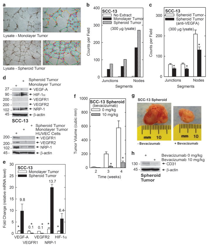
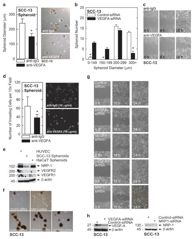
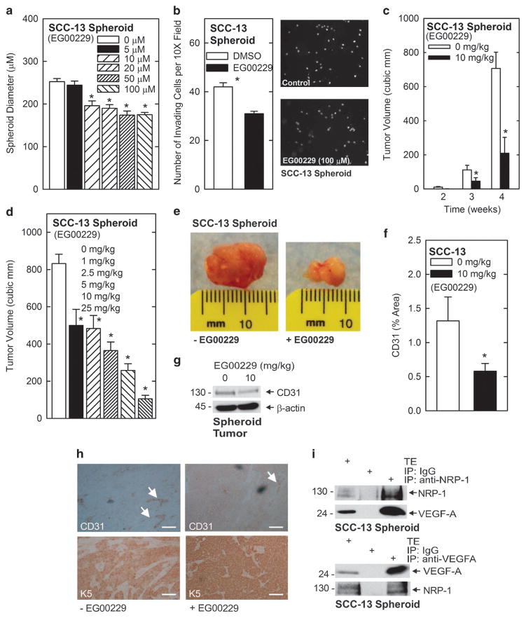
We identify a limited subpopulation of epidermal cancer stem cells (ECS cells), in squamous cell carcinoma, that form rapidly growing, invasive and highly vascularized tumors, as compared with non-stem cancer cells. These ECS cells grow as non-attached spheroids, and display enhanced migration and invasion. We show that ECS cell-produced vascular endothelial growth factor (VEGF)-A is required for the maintenance of this phenotype, as knockdown of VEGF-A gene expression or treatment with VEGF-A-inactivating antibody reduces these responses. In addition, treatment with bevacizumab reduces tumor vascularity and growth. Surprisingly, the classical mechanism of VEGF-A action via interaction with VEGF receptors does not mediate these events, as these cells lack VEGFR1 and VEGFR2. Instead, VEGF-A acts via the neuropilin-1 (NRP-1) co-receptor. Knockdown of NRP-1 inhibits ECS cell spheroid formation, invasion and migration, and attenuates tumor formation. These studies suggest that VEGF-A acts via interaction with NRP-1 to trigger intracellular events leading to ECS cell survival and formation of aggressive, invasive and highly vascularized tumors.


Conflict of interest statement

The authors declare no conflict of interest.

Figures

Figure 1
Figure 1
Epidermal cancer stem (ECS) cells form large and highly vascularized tumors. (a) Images of SCC-13 cells grown as monolayer (attached) or spheroid (unattached) cultures. (b) Stem marker expression in monolayer cells versus spheroids. Cells were grown for 10 days and total extracts were prepared for immunoblot detection of the indicated proteins. Similar results were observed in each of three independent experiments. (c, d) Tumor morphology and histology. Non-stem cancer cells and ECS cells (0.1 million/injection site) were injected in each front flank in NSG mice. After 4 weeks, the tumors were harvested and photographed. Primary tumors were paraffin-embedded, sectioned and stained with H&E, or antibodies detecting CD31 (endothelial cells) and K5 (epithelial cells). The arrows indicate blood vessels, and the bars =125 microns. (e) ImageJ quantification of CD31 distribution. CD31 distribution was quantified using ImageJ and expressed as a percentage of the total area. The values are mean ± s.e.m., n =6, and the asterisks indicate a significant difference, P<0.05. The arrows indicate CD31 staining. (f) CD31 level. Tumor extracts from non-stem cancer (monolayer) and ECS cell (spheroid)-derived tumors were electrophoresed for immunoblot detection of CD31. (g) K5 and human nuclear antigen (HNA) distribution. The arrow shows HNA- and K5-positive human epithelial tumor cells. Note that the mesenchymal cells, which include the blood vessels, are K5 and HNA negative.
Figure 2
Figure 2
VEGF-A angiogenic activity in ECS cell-derived tumors. (a) ECS cell tumor extracts stimulate vessel formation. Extracts were prepared from non-stem cancer cell- or ECS cell-derived tumors and 300 μg of lysate was incubated with HUVEC cultures for 18 h. Images were collected (left panels) and analyzed for detection of junction, segment and node formation (right panels). (b) Plot showing frequency of junctions, segments and nodes detected by ImageJ analysis of images in panel a. (c) Anti-VEGF-A inhibits vessel formation. ECS cell tumor lysate (300 μg) was treated with 0 or 10 μg/ml anti-VEGF-A and then tested in a HUVEC assay for ability to stimulate vessel formation. The values are mean ± s.e.m., n =3, and the asterisk indicates a significant difference, P<0.05. (d) VEGF signaling protein expression in tumors. Tumor extracts were prepared for immunoblot detection of the indicated proteins. (e) Level of mRNA-encoding VEGF signaling protein in tumors. RNA was isolated from non-stem cancer- and ECS cell-derived tumors for qRT-PCR measurement of mRNA-encoding angiogenic signaling proteins. The values are mean ± s.e.m., n = 3, and the asterisks indicate a significant difference, P<0.05. (f, g) Bevacizumab suppresses tumor formation. ECS cells (0.1 million) were injected into the two front flanks of NSG mice, treatment was initiated with 0 or 10 mg/kg bevacizumab and tumor size and morphology was monitored. The values are mean ± s.e.m., n = 6, and the asterisks indicate a significant difference, P<0.05. The tumor images are from 4 week tumors. (h) Immunoblot detection of CD31 in ECS cell tumors. ECS cells (0.1 million) were injected into each front flank of NSG mice, and bevacizumab was delivered by intraperitoneal injection three times per week at a level of 10 mg/kg body weight. After 4 weeks the tumors were harvested and extracts were prepared for detection of CD31.
Figure 3
Figure 3
VEGF-A signaling proteins and control of ECS cell function. (a, b) Anti-VEGF-A impact on spheroid formation. SCC-13 cells were seeded at 40 000 cells/well in ultra-low attachment plates in spheroid medium containing 10 μg/ml anti-VEGF or control IgG. After 3 days, spheroid number, and size distribution, were analyzed and expressed as mean ± s.e.m., n = 4, and the asterisks indicate a significant difference, P<0.05. (c, d) Anti-VEGF-A treatment suppresses ECS cell migration and invasion. ECS cells were plated at confluent density on conventional culture plates and then scratched to create uniform wounds. Anti-VEGF-A (0 or 10 μg/ml) was added at the time of wounding and wound width was monitored at 0, 8 and 18 h. ECS cells were seeded into a transwell chamber atop a matrigel layer in the presence of 0 or 10 μg/ml anti-VEGF-A and cell invasion was monitored at 18 h. The asterisks indicate a significant difference, n = 3, P<0.05. (e) VEGF-A signaling protein expression. Extracts were prepared from monolayer cultures of the indicated cell lines for immunoblot detection of the indicated proteins. (f, g) VEGF-A and NRP-1 are required for ECS cell spheroid formation and migration. SCC-13 monolayer cells were electroporated with 3 μg of control-, VEGF-A-, VEGFR1-, VEGFR2- or NRP-1-siRNA and plated in triplicate in ultra-low attachment dishes at 40 000 cells per well and spheroids were photographed after 5 days. In parallel, the electroporated cells were seeded at confluence in conventional plates, permitted to attach and uniformly wounded using a 10 μl pipet tip. Spheroid medium was added and wound closure was monitored at 0, 16 and 24 h. (h) Immunoblot confirmation of VEGF-A and NRP-1 knockdown in siRNA-treated ECS cells used in panels f and g. We routinely achieve a near-complete reduction in VEGF-A level and a 50% reduction in NRP-1 level.
Figure 4
Figure 4
EG00229 suppresses ECS cell function and tumor formation. (a) EG00229 impact on ECS cell spheroid formation. SCC-13 cells were seeded at 40 000 cells per well in ultra-low attachment plates in spheroid medium containing 0–100 μM EG00229. After 3 days, spheroid number was analyzed and expressed as mean ± s.e.m., n = 3, P<0.05. (b) EG00229 impact on ECS cell matrigel invasion. ECS cells were seeded into a transwell chambers atop a matrigel layer in the presence of 0 or 100 μM EG00229 and invasion was monitored at 18 h. The values are mean ±s.e.m. and the asterisk indicates a significant difference, n = 3, P<0.05. (ch) EG00229 suppresses tumor formation and vascularization. ECS cells were enriched by growth for 10 days as spheroids, and 0.1 million cells were injected into NSG mice in each front flank. The mice were treated with 0 or 10 mg EG00229/kg body weight for various times (c) or various doses of EG00229 for 4 week (d) given three times per week by IP injection. Tumor formation was monitored using calipers and tumors were photographed on week 4 at the time of harvest (e, 10 mg/kg body weight). The values are mean ± s.e.m., n =6, and the asterisks indicates a significant difference, P<0.05. EG00229 treatment did not impact animal weight. Sections were stained with anti-CD31 and processed by ImageJ and staining intensity was expressed as % area (f). The values are mean ± s.e.m., n =6, and the asterisk indicates a significant difference, P<0.05. The reduction in CD31 level was confirmed by immunoblot of tumor extracts (g). Similar results were observed for each of three tumor comparisons. Tumor sections were prepared from tumors treated with 0 or 10 mg/kg body weight EG00229 and then stained to detect CD31 (vascularization marker) and keratin 5 (K5, epithelial cell marker) (h). The arrows indicate CD31-positive blood vessels. The bar =125 μm. (i) Evidence for NRP-1/VEGF-A interaction. Extracts were prepared from SCC-13-derived ECS cells in lysis buffer and 200 μg of extract was immunoprecipitated with anti-NRP-1 or anti-VEGF-A followed by immunoblot to detect the indicated epitopes. Total extract (25 μg) was electrophoresed as a control. Similar results were observed in three experiments.

References

    1. American Cancer Society Website. Cancer Facts and Figures. 2010 Available at: http://www.cancer.org/
    1. Bickers DR, Lim HW, Margolis D, Weinstock MA, Goodman C, Faulkner E, et al. The burden of skin diseases: 2004 a joint project of the American Academy of Dermatology Association and the Society for Investigative Dermatology. J Am Acad Dermatol. 2006;55:490–500. - PubMed
    1. Rogers HW, Weinstock MA, Harris AR, Hinckley MR, Feldman SR, Fleischer AB, et al. Incidence estimate of nonmelanoma skin cancer in the United States, 2006. Arch Dermatol. 2010;146:283–287. - PubMed
    1. Pleasance ED, Cheetham RK, Stephens PJ, McBride DJ, Humphray SJ, Greenman CD, et al. A comprehensive catalogue of somatic mutations from a human cancer genome. Nature. 2010;463:191–196. - PMC - PubMed
    1. Folkman J. The role of angiogenesis in tumor growth. Semin Cancer Biol. 1992;3:65–71. - PubMed

Publication types

MeSH terms

Substances