. 2016 Apr:38:103-110.
doi: 10.1016/j.anaerobe.2016.01.004. Epub 2016 Jan 21.

A novel murine model of Clostridium sordellii myonecrosis: Insights into the pathogenesis of disease

Affiliations

A novel murine model of Clostridium sordellii myonecrosis: Insights into the pathogenesis of disease

Michael J Aldape et al. Anaerobe. 2016 Apr.

Abstract

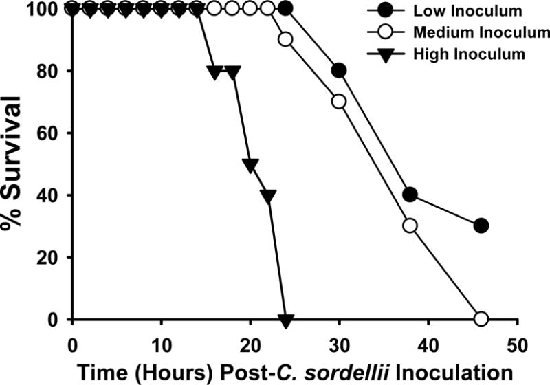
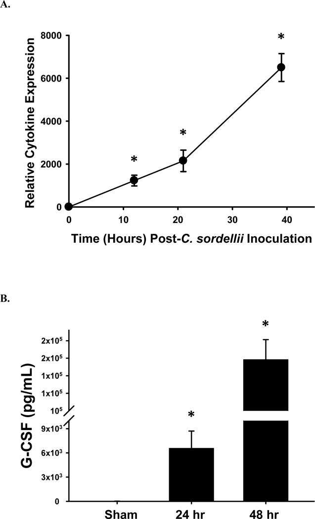
Clostridium sordellii infections have been reported in women following natural childbirth and spontaneous or medically-induced abortion, injection drug users and patients with trauma. Death is rapid and mortality ranges from 70 to 100%. Clinical features include an extreme leukemoid reaction, the absence of fever, and only minimal pain or erythema at the infected site. In the current study, we developed a murine model of C. sordellii soft tissue infection to elucidate the pathogenic mechanisms. Mice received 0.5, 1.0 or 2.0 × 10(6) CFU C. sordellii (ATCC 9714 type strain) in the right thigh muscle. All doses caused fatal infection characterized by intense swelling of the infected limb but no erythema or visible perfusion deficits. Survival rates and time to death were inoculum dose-dependent. Mice developed a granulocytic leukocytosis with left shift, the onset of which directly correlated with disease severity. Histopathology of infected tissue showed widespread edema, moderate muscle damage and minimal neutrophil infiltration. Circulating levels of granulocyte colony-stimulating factor (G-CSF), soluble tumor necrosis factor receptor I (sTNF-RI) and interlukin-6 (IL-6) were significantly increased in infected animals, while TNF-α, and IL-1β levels were only mildly elevated, suggesting these host factors likely mediate the leukocytosis and innate immune dysfunction characteristic of this infection. Thus, this model mimics many of the salient features of this infection in humans and has allowed us to identify novel targets for intervention.

Keywords: C. sordellii; Host immune response; Leukemoid reaction; Myonecrosis; Pathogenesis.


Conflict of interest statement

Conflict of interest: No authors involved with this manuscript have a commercial or other association that might pose a conflict of interest.

Figures

Figure 1
Figure 1. Survival curves of mice infected intramuscularly with C. sordellii ATCC 9714
Groups of C57BL/6 mice (N = 10) were inoculated in the right thigh muscle with either 0.5 (low), 1.0 (medium) or 2.0 (high) × 106 colony forming units of washed, log phase C. sordellii ATCC 9714. Mice were observed for 5 days with mortalities recorded.
Figure 2
Figure 2. Comparative physical findings of mice infected intramuscularly with C. sordellii (A), or control (B) versus C. perfringens (C)
C57BL/6 mice were inoculated with 1.0×10⁶ CFU of washed, log phase C. sordellii ATCC 9714 (A). C. sordellii infected animals developed severe edema at the infection site compared to non-infected mice (A vs B) but little erythema of the muscle or overlying skin (not shown) or blackening of the foot that are characteristic of experimental C. perfringens infection (A vs C).
Figure 3
Figure 3. C. sordellii muscle infection causes marked histologic abnormalities
Routine hematoxylin-eosin staining of muscle specimens obtained from 4 mice 39 hrs after infection with 1.0×10⁶ CFU C. sordellii ATCC 9714; control mice received sterile saline injection. (A) H&E of normal muscle tissue at 20× magnification. H&E of infected muscle at (B) 20× and (C) 40× magnification shows widespread edema and areas of focal PMNL influx and tissue necrosis. (D) Muscle specimens from infected animals were scored by a blinded observer for evidence of inflammation, edema, tissue necrosis and hemorrhage on a scale of 0–3 for each abnormality (max score 12). No pathologies were observed in sham-infected control muscles (not shown).
Figure 4
Figure 4. Intramuscular infection with varying doses of C. sordellii generates a leukemoid reaction
C57BL/6 mice were infected intramuscularly with either, 0.5 (low), 1.0 (medium) or 2.0 (high) × 106 colony forming units of washed, log phase C. sordellii ATCC 9714. (A) Total WBC counts were obtained on blood specimens from 2 randomly chosen mice at that time point. Data are the mean maximal counts (± standard deviation) observed for each inoculum group. Sham infected mice had a constant WBC count of 4.2 × 10³/μL whole blood. WBC differential ratios were determined by a blinded observer with expertise in this area. (B) Polymorphonuclear leukocytes, (C) Granulocyte precursors, and (D) Lymphocytes.
Fig. 5
Fig. 5. Granulocyte-colony stimulating factor response in mice infected intramuscularly with C. sordellii
(A) Fold change differences in circulating G-CSF levels in mice infected with 1 × 10⁶ CFU of C. sordellii ATCC 9714 as determined by murine cytokine array analysis. Serum was collected from two infected mice at each indicated time. (B) Serum concentrations of G-CSF as determined by ELISA. Data represents the average serum concentration from four mice infected with either nothing or 1 × 10⁶ CFU of C. sordellii at 24 hrs and 48 hrs. *P< .05, compared with the no-toxin (NoTx) control (1-way analysis of variance with Tukey’s test).
Fig. 6
Fig. 6. Serum concentrations of innate immune modulators from mice infected intramuscularly with C. sordellii
(A) Relative fold change differences in circulating TNF-α, IL-1β and IL-6 in mice infected with 1 × 10⁶ CFU of C. sordellii ATCC 9714 as determined by murine cytokine array analysis. Serum was collected from two infected mice at each indicated time. (B) Serum concentrations of sTNF-R1 as determined by ELISA. Data represents the average serum concentration from four mice challenged with either nothing or 1 × 10⁶ CFU of C. sordellii at 24 hrs and 48 hrs post-infection. *P< .05, compared with the no-toxin (NoTx) control (1-way analysis of variance with Tukey’s test).

References

    1. Aldape MJ, Bryant AE, Stevens DL. Clostridium sordellii infection: epidemiology, clinical findings, and current perspectives on diagnosis and treatment. Clin Infect Dis. 2006 Dec 1;43(11):1436–46. - PubMed
    1. Sicard D. Deaths from Clostridium sordellii after medical abortion. N Engl J Med. 2006 Apr 13;354(15):1645–7. - PubMed
    1. Sinave C, Le TG, Blouin D, Leveille F, Deland E. Toxic shock syndrome due to Clostridium sordellii: a dramatic postpartum and postabortion disease. Clin Infect Dis. 2002 Dec 1;35(11):1441–3. - PubMed
    1. Smith LDS. Clostridium sordellii. In: Smith LDS, editor. The pathogenic anaerobic bacteria. Springfield, IL: Charles C. Thomas Publishing; 1975. pp. 291–8.
    1. Aronoff DM, Hao Y, Chung J, et al. Misoprostol impairs female reproductive tract innate immunity against Clostridium sordellii. J Immunol. 2008 Jun 15;180(12):8222–30. - PMC - PubMed

Publication types

LinkOut - more resources