

Aging alters bone-fat reciprocity by shifting in vivo mesenchymal precursor cell fate towards an adipogenic lineage
- PMID: 26805026
- PMCID: PMC4792752
- DOI: 10.1016/j.bone.2016.01.014
Aging alters bone-fat reciprocity by shifting in vivo mesenchymal precursor cell fate towards an adipogenic lineage
Abstract
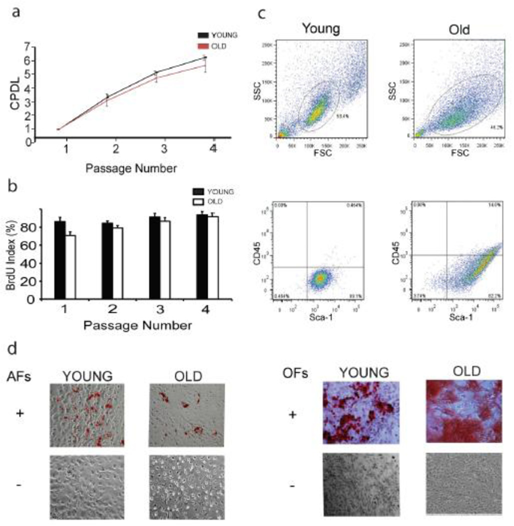
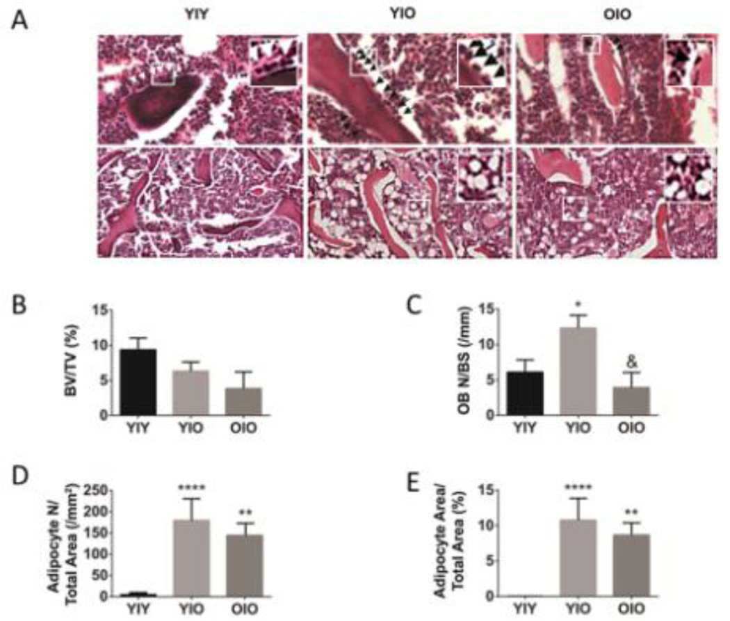
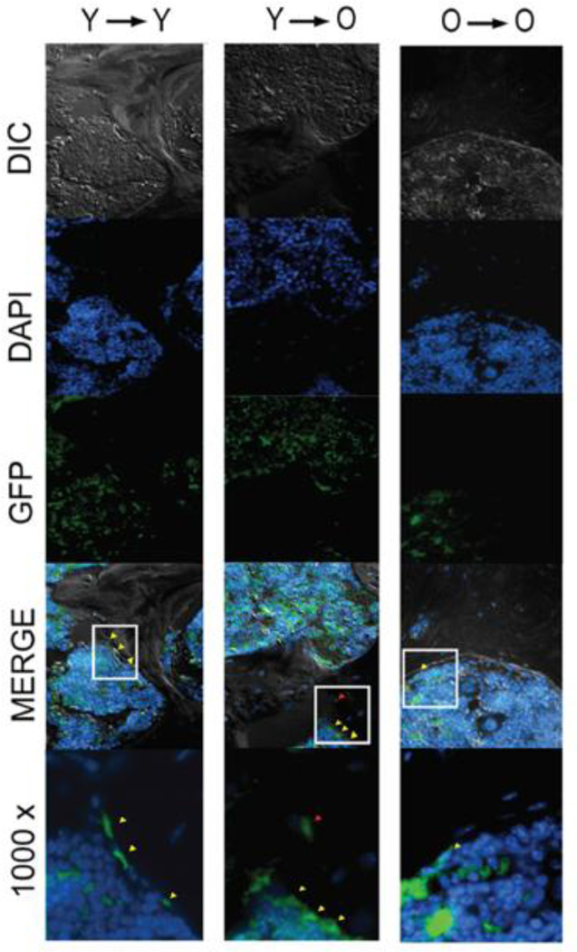
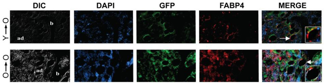
Bone marrow derived mesenchymal progenitor cells (MPCs) play an important role in bone homeostasis. Age-related changes occur in bone resulting in a decrease in bone density and a relative increase in adipocity. Although in vitro studies suggest the existence of an age-related lineage switch between osteogenic and adipogenic fates, stem cell and microenvironmental contributions to this process have not been elucidated in vivo. In order to study the effects of MPC and microenvironmental aging on functional engraftment and lineage switching, transplantation studies were performed under non-myeloablative conditions in old recipients, with donor MPCs derived from young and old green fluorescent protein (GFP) transgenic mice. Robust engraftment by young MPCs or their progeny was observed in the marrow, bone-lining region and in the matrix of young recipients; however, significantly lower engraftment was seen at the same sites in old recipients transplanted with old MPCs. Differentiation of transplanted MPCs strongly favored adipogenesis over osteogenesis in old recipients irrespective of MPC donor age, suggesting that microenvironmental alterations that occur with in vivo aging are predominately responsible for MPC lineage switching. These data indicate that aging alters bone-fat reciprocity and differentiation of mesenchymal progenitors towards an adipogenic fate.
Keywords: Aging; Bone; Engraftment; Fat; Mesenchymal stem cells; Osteoporosis.
Copyright © 2016 Elsevier Inc. All rights reserved.
Conflict of interest statement
Figures
References
-
- Burkhardt R, Kettner G, Bohm W, Schmidmeier M, Schlag R, Frisch B, Mallmann B, Eisenmenger W, Gilg T. Changes in trabecular bone, hematopoiesis and bone marrow vessels in aplastic anemia, primary osteoporosis, and old age: a comparative histomorphometric study. Bone. 1987;8:157–164. - PubMed
-
- Rozman C, Reverter JC, Feliu E, Berga L, Rozman M, Climent C. Variations of fat tissue fraction in abnormal human bone marrow depend both on size and number of adipocytes: a stereologic study. Blood. 1990;76:892–895. - PubMed
-
- Justesen J, Stenderup K, Ebbesen EN, Mosekilde L, Steiniche T, Kassem M. Adipocyte tissue volume in bone marrow is increased with aging and in patients with osteoporosis. Biogerontology. 2001;2:165–171. - PubMed
-
- Meunier P, Aaron J, Edouard C, Vignon G. Osteoporosis the replacement of cell populations of the marrow by adipose tissue. A quantitative study of 84 iliac bone biopsies. Clin Orthop Relat Res. 1971;80:147–154. - PubMed
-
- Minaire P, Neunier P, Edouard C, Bernard J, Courpron P, Bourret J. Quantitative histological data on disuse osteoporosis: comparison with biological data. Calcif Tissue Res. 1974;17:57–73. - PubMed
Publication types
MeSH terms
Grants and funding
LinkOut - more resources
Full Text Sources
Other Literature Sources
Medical
