Review
. 2016 Jan;53(1):10-9.
doi: 10.1053/j.seminhematol.2015.10.005. Epub 2015 Oct 26.

Advances and challenges in hemophilic arthropathy

Affiliations
Review

Advances and challenges in hemophilic arthropathy

Tine Wyseure et al. Semin Hematol. 2016 Jan.

Abstract

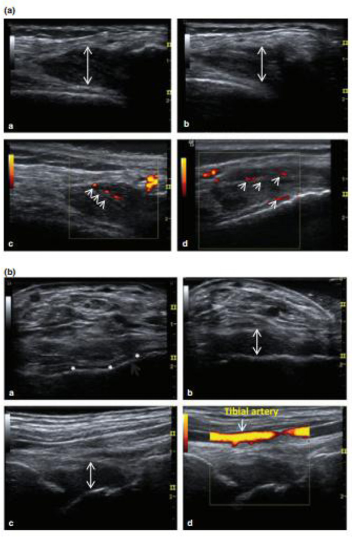
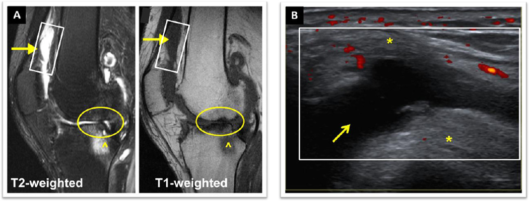
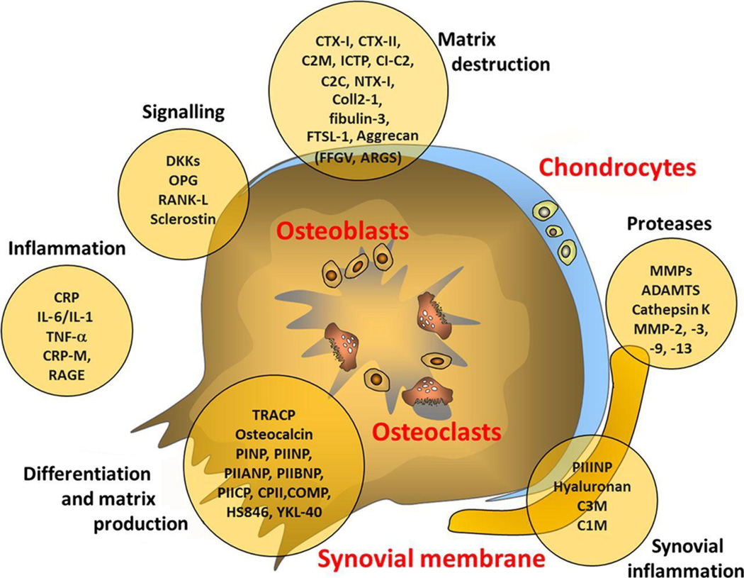
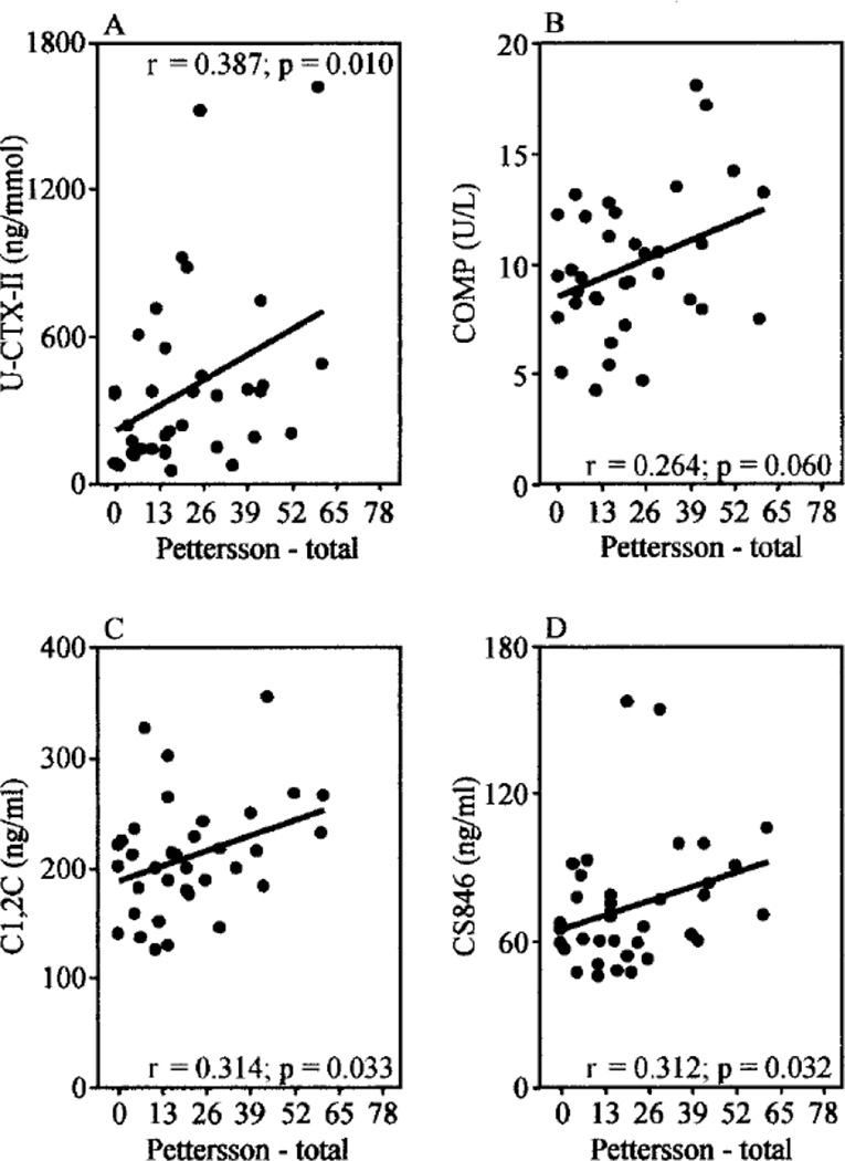
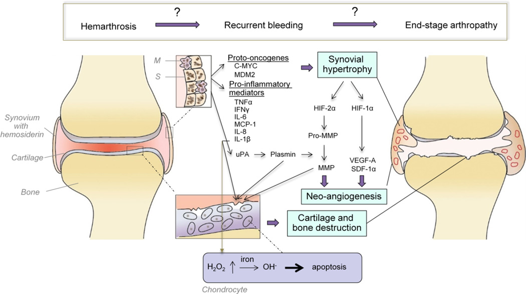
Hemophilic arthropathy is a form of joint disease that develops secondary to joint bleeding and presents with synovial hypertrophy, cartilage and bony destruction. The arthropathy can develop despite clotting factor replacement and is especially disabling in the aging population. Pathobiological tissue changes are triggered by release of hemoglobin and iron deposition in the joint, but the sequence of events and the molecular mechanisms resulting in joint deterioration are incompletely understood. Treatment options other than clotting factor replacement are limited. Improvements in the treatment of hemophilia necessitate a better understanding of the processes that lead to this disabling condition and better diagnostic tools. Towards that end, studies of the molecular mechanisms leading to the arthropathy, as well as the development of sensitive imaging techniques and biomarkers are needed. These will pave the way to identify the cause of acute pain such as joint bleeding or synovitis, detect early, potentially reversible structural changes, and predict progression of disease. This review describes current imaging techniques and the development of high resolution musculoskeletal ultrasound with power Doppler to afford point-of-care diagnosis and management, the potential utility of diagnostic biomarkers, and summarizes our current knowledge of the pathobiology of hemophilic arthropathy.

Keywords: Arthropathy; Biomarkers; Hemarthrosis; Hemophilia; Imaging.


Figures

Figure 1
Figure 1. Two case studies illustrating musculoskeletal ultrasound (MSKUS) in the diagnosis of bleeding and non-bleeding episodes during acute painful episodes
(A) 37-year-old man with severe haemophilia A presented with perceived joint bleeding. a: Axial view of the lateral gutter of the left knee showed heterogeneous hypoechoic intra-articular material (arrow). b: The intra-articular (i/a) material was hardly compressible and not displaceable (arrow) suggestive of thickened synovial tissue. c/d: power Doppler (PD) revealed vascularization of thickened hypervascular synovial tissue without evidence of sonographic effusion. This episode was reclassified as synovitis. (B) 23-year-old man with severe haemophilia B presented with ankle pain and perceived “arthritis pain”. a: Baseline axial view of the ankle showed thin anechoic joint space in the tibiotalar joint (*). b: Interval examination during pain episode showed marked volume increased tibiotalar synovial space (arrow). Complex echogenic pattern and compressibility of the synovial space were consistent with complex effusion//intra-articular bleed. c/d: Orthogonal view without and with power Doppler demonstrated absence of tissue vascularization. This episode was reclassified as acute haemarthrosis Ceponis, A. et al. Haemophilia 2013. 19: pp 790–798. Modified. With permission form the journal.
Figure 2
Figure 2. Comparison of MRI and MSKUS of the knee of a 54 year old man with moderate Hemophilia A. Both imaging studies were performed on the same day
The patient presented with persistent knee swelling and pain 2 weeks after a fall. A) MRI (sagittal view): ^ Marrow edema, O cartilage and meniscus destruction. The arrows (→) indicate areas in the suprapatellar bursa and patellar recesses where diagnostic distinctions of synovial hypertrophy/synovitis from effusion, and of bloody from non-bloody effusions were difficult by conventional MRI. The rectangle depicts the same area scanned with B) MSKUS. MSKUS demonstrated a simple non-bloody effusion (anechoic, compressible area (→)) surrounded by *fat pad and *synovium. Power Doppler (red dots) revealed areas of synovial perfusion. MRI, Magnet Resonance Imaging. MSKUS, Musculoskeletal Ultrasound. University of California San Diego Case Series, Images provided by Annette von Drygalski, MD. Patient provided written informed consent.
Figure 3
Figure 3. Sources of possible biomarkers in osteoarthritis
10–20 C2C, cleavage of type II collagen; C2M, collagen type II-specific neoepitope; C3M, collagen type III-specific neoepitope; Coll 2-1, 9-amino acid peptide of type II collagen (nitrated form Coll 2-1 NO2); COMP, cartilage oligomeric protein; CPII, type II collagen propeptide; CRP, C reactive protein; CTX, C-terminal telopeptide of collagen; DKK, wnt inhibitor; FSTL-1, follistatin-like protein 1; ICTP, type I collagen-derived cross-linked carboxy-terminal telopeptide; IL, interleukin; MMP, matrix metalloproteinase; NTX, N-terminal telopeptide of collagen OPG, osteoprotegerin; PIIANP, N-propeptide IIA of type II collagen; PIIBNP, N-propeptide IIB of type II collagen; PIICP, C-propeptide of collagen type II; PINP, N-propeptide of type I collagen; PIINP, N-propeptide of type II collagen; RANK-L, receptor activator of nuclear factor κB ligand; RAGE, receptor for advanced glycation endproducts; TNF, tumour necrosis factor. Lotz M, et al. Postgrad Med J 2014; 90: pp 171–178. With permission from the Journal.
Figure 4
Figure 4
The correlation of radiographic joint damage with biomarker levels of cartilage turn-over with radiographic Pettersson scores in hemophilic arthropathy. Jansen N.W.D., et al. Arthritis & Rheumatism 2009; 60: pp 290–298. With permission from the Journal.
Figure 5
Figure 5. Pathobiology of hemophilic arthropathy. A provisional scheme with many open questions
Erythrocytes in the joint space are taken up by synoviocytes (S) and tissue macrophages (M), that store iron released from erythrocytes as hemosiderin. The expression of the oncoproteins C-MYC and MDM2 in response to iron results in synovial hypertrophy. Synovial hypertrophy creates a hypoxic environment causing a rise in hypoxia inducible factor (HIF)-1α and HIF-2α, which induce increased expression of the pro-angiogenic mediators vascular endothelial growth factor-A (VEGF-A) and stromal-cell derived factor 1α (SDF-1α), as well as pro-matrix metalloproteinases (pro-MMP). VEGF-A stimulates synovial neoangiogenesis and plasmin-mediated conversion to MMPs results in glycosaminoglycan release from the cartilage matrix, and cartilage and subchondral bone destruction. Synoviocytes and tissue macrophages also release pro-inflammatory mediators, such as tumor necrosis factor alpha (TNFα), interferon-γ, interleukin (IL)-1β, IL-6, monocyte chemoattractant protein-1 (MCP-1) fueling these processes. Secreted IL-1β induces increased production of hydrogen peroxide (H2O2) in the chondrocytes, which upon reaction with iron (Fe2+) forms cytotoxic hydroxyl radicals (OH) and prompts chondrocyte apoptosis. Neoangiogenesis and vascular remodeling are assumed to maintain a vicious cycle of re-bleeding that results in progressive hemophilic arthropathy, characterized by synovial hypertrophy, cartilage and bone destruction and joint deformities.

References

    1. Berntorp E, Shapiro AD. Modern haemophilia care. Lancet. 2012;379:1447–1456. - PubMed
    1. Soucie JM, Cianfrini C, Janco RL, et al. Joint range-of-motion limitations among young males with hemophilia: prevalence and risk factors. Blood. 2004;103:2467–2473. - PubMed
    1. Aledort LM, Haschmeyer RH, Pettersson H. A longitudinal study of orthopaedic outcomes for severe factor-VIII-deficient haemophiliacs. The Orthopaedic Outcome Study Group. J Intern Med. 1994;236:391–399. - PubMed
    1. Oldenburg J, Zimmermann R, Katsarou O, et al. Controlled, cross-sectional MRI evaluation of joint status in severe haemophilia A patients treated with prophylaxis vs. on demand. Haemophilia. 2015;21:171–179. - PMC - PubMed
    1. Fischer K, Steen Carlsson K, Petrini P, et al. Intermediate-dose versus high-dose prophylaxis for severe hemophilia: comparing outcome and costs since the 1970s. Blood. 2013;122:1129–1136. - PMC - PubMed

Publication types