

Abnormal brain activation and connectivity to standardized disorder-related visual scenes in social anxiety disorder
- PMID: 26806013
- PMCID: PMC6867294
- DOI: 10.1002/hbm.23120
Abnormal brain activation and connectivity to standardized disorder-related visual scenes in social anxiety disorder
Abstract
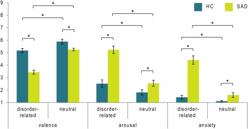
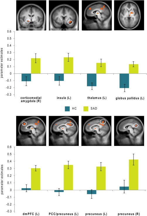
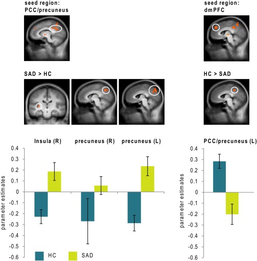
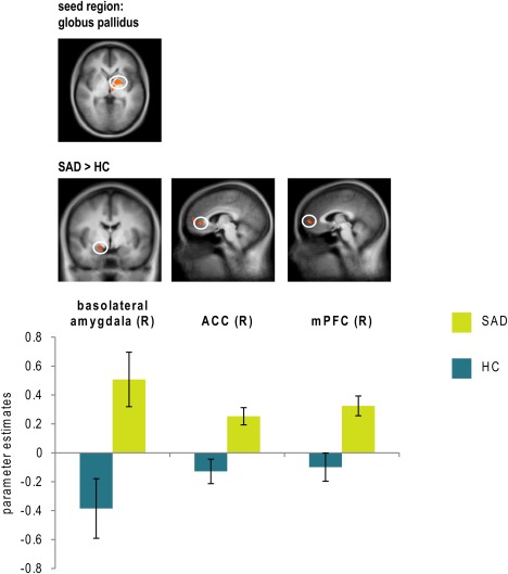
Our understanding of altered emotional processing in social anxiety disorder (SAD) is hampered by a heterogeneity of findings, which is probably due to the vastly different methods and materials used so far. This is why the present functional magnetic resonance imaging (fMRI) study investigated immediate disorder-related threat processing in 30 SAD patients and 30 healthy controls (HC) with a novel, standardized set of highly ecologically valid, disorder-related complex visual scenes. SAD patients rated disorder-related as compared with neutral scenes as more unpleasant, arousing and anxiety-inducing than HC. On the neural level, disorder-related as compared with neutral scenes evoked differential responses in SAD patients in a widespread emotion processing network including (para-)limbic structures (e.g. amygdala, insula, thalamus, globus pallidus) and cortical regions (e.g. dorsomedial prefrontal cortex (dmPFC), posterior cingulate cortex (PCC), and precuneus). Functional connectivity analysis yielded an altered interplay between PCC/precuneus and paralimbic (insula) as well as cortical regions (dmPFC, precuneus) in SAD patients, which emphasizes a central role for PCC/precuneus in disorder-related scene processing. Hyperconnectivity of globus pallidus with amygdala, anterior cingulate cortex (ACC) and medial prefrontal cortex (mPFC) additionally underlines the relevance of this region in socially anxious threat processing. Our findings stress the importance of specific disorder-related stimuli for the investigation of altered emotion processing in SAD. Disorder-related threat processing in SAD reveals anomalies at multiple stages of emotion processing which may be linked to increased anxiety and to dysfunctionally elevated levels of self-referential processing reported in previous studies.
Keywords: amygdala; default-mode-network; globus pallidus; precuneus; psychophysiological interaction; self-referential processing; social phobia.
© 2016 Wiley Periodicals, Inc.
Figures
Similar articles
-
Brain activation to task-irrelevant disorder-related threat in social anxiety disorder: The impact of symptom severity.Neuroimage Clin. 2017 Jan 23;14:323-333. doi: 10.1016/j.nicl.2017.01.020. eCollection 2017. Neuroimage Clin. 2017. PMID: 28224080 Free PMC article.
-
Network analysis reveals disrupted functional brain circuitry in drug-naive social anxiety disorder.Neuroimage. 2019 Apr 15;190:213-223. doi: 10.1016/j.neuroimage.2017.12.011. Epub 2017 Dec 7. Neuroimage. 2019. PMID: 29223742
-
Resting-State Functional Connectivity in Generalized Anxiety Disorder and Social Anxiety Disorder: Evidence for a Dimensional Approach.Brain Connect. 2017 Jun;7(5):289-298. doi: 10.1089/brain.2017.0497. Brain Connect. 2017. PMID: 28478685
-
Beyond emotions: A meta-analysis of neural response within face processing system in social anxiety.Exp Biol Med (Maywood). 2016 Feb;241(3):225-37. doi: 10.1177/1535370215603514. Epub 2015 Sep 3. Exp Biol Med (Maywood). 2016. PMID: 26341469 Free PMC article. Review.
-
Topography of the Anxious Self: Abnormal Rest-Task Modulation in Social Anxiety Disorder.Neuroscientist. 2023 Apr;29(2):221-244. doi: 10.1177/10738584211030497. Epub 2021 Jul 20. Neuroscientist. 2023. PMID: 34282680 Review.
Cited by
-
Gray Matter Structural Alterations in Social Anxiety Disorder: A Voxel-Based Meta-Analysis.Front Psychiatry. 2018 Sep 21;9:449. doi: 10.3389/fpsyt.2018.00449. eCollection 2018. Front Psychiatry. 2018. PMID: 30298028 Free PMC article. Review.
-
Increased default mode network activation in depression and social anxiety during upward social comparison.Soc Cogn Affect Neurosci. 2025 Feb 4;20(1):nsaf012. doi: 10.1093/scan/nsaf012. Soc Cogn Affect Neurosci. 2025. PMID: 39882939 Free PMC article.
-
Imaging the socially-anxious brain: recent advances and future prospects.F1000Res. 2020 Apr 2;9:F1000 Faculty Rev-230. doi: 10.12688/f1000research.21214.1. eCollection 2020. F1000Res. 2020. PMID: 32269760 Free PMC article. Review.
-
Intrinsic functional connectivity in families genetically enriched for social anxiety disorder - an endophenotype study.EBioMedicine. 2021 Jul;69:103445. doi: 10.1016/j.ebiom.2021.103445. Epub 2021 Jun 20. EBioMedicine. 2021. PMID: 34161885 Free PMC article.
-
Engagement and Disengagement: From the Basic Science of Emotion Regulation to an Anxiety Spectrum.Psychophysiology. 2025 Feb;62(2):e70006. doi: 10.1111/psyp.70006. Psychophysiology. 2025. PMID: 39924448 Free PMC article. Review.
References
-
- Alexander GE, Crutcher MD (1990): Functional architecture of basal ganglia circuits: Neural substrates of parallel processing. Trends Neurosci 13:266–271. - PubMed
-
- American Psychiatric Association (2000): Diagnostic and Statistical Manual of Mental Disorders, Fourth Edition: DSM‐IV‐TR. Washington, DC: American Psychiatric Publications.
-
- Amir N, Klumpp H, Elias J, Bedwell JS, Yanasak N, Miller LS (2005): Increased activation of the anterior cingulate cortex during processing of disgust faces in individuals with social phobia. Biol Psychiatry 57:975–981. - PubMed
-
- Amunts K, Kedo O, Kindler M, Pieperhoff P, Mohlberg H, Shah NJ, Habel U, Schneider F, Zilles K (2005): Cytoarchitectonic mapping of the human amygdala, hippocampal region and entorhinal cortex: Intersubject variability and probability maps. Anat Embryol (Berl) 210:343–352. - PubMed
-
- Binelli C, Subirà S, Batalla A, Muñiz A, Sugranyés G, Crippa JA, Farré M, Pérez‐Jurado L, Martín‐Santos R (2014): Common and distinct neural correlates of facial emotion processing in social anxiety disorder and Williams syndrome: A systematic review and voxel‐based meta‐analysis of functional resonance imaging studies. Neuropsychologia 64:205–217. - PubMed
Publication types
MeSH terms
LinkOut - more resources
Full Text Sources
Other Literature Sources
Medical
