. 2016 May;194(2):139-46.
doi: 10.1016/j.jsb.2016.01.006. Epub 2016 Jan 13.

Tectorins crosslink type II collagen fibrils and connect the tectorial membrane to the spiral limbus

Affiliations

Tectorins crosslink type II collagen fibrils and connect the tectorial membrane to the spiral limbus

Leonardo R Andrade et al. J Struct Biol. 2016 May.

Abstract

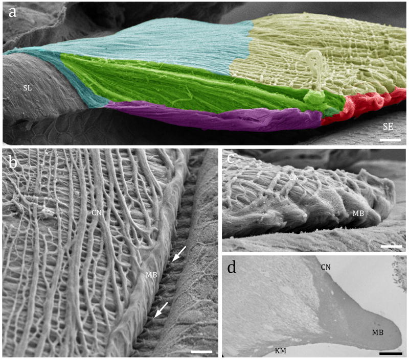
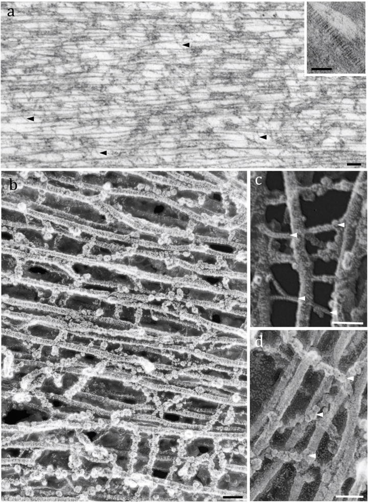
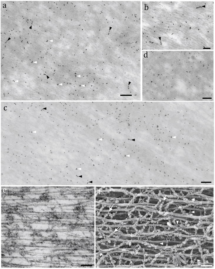
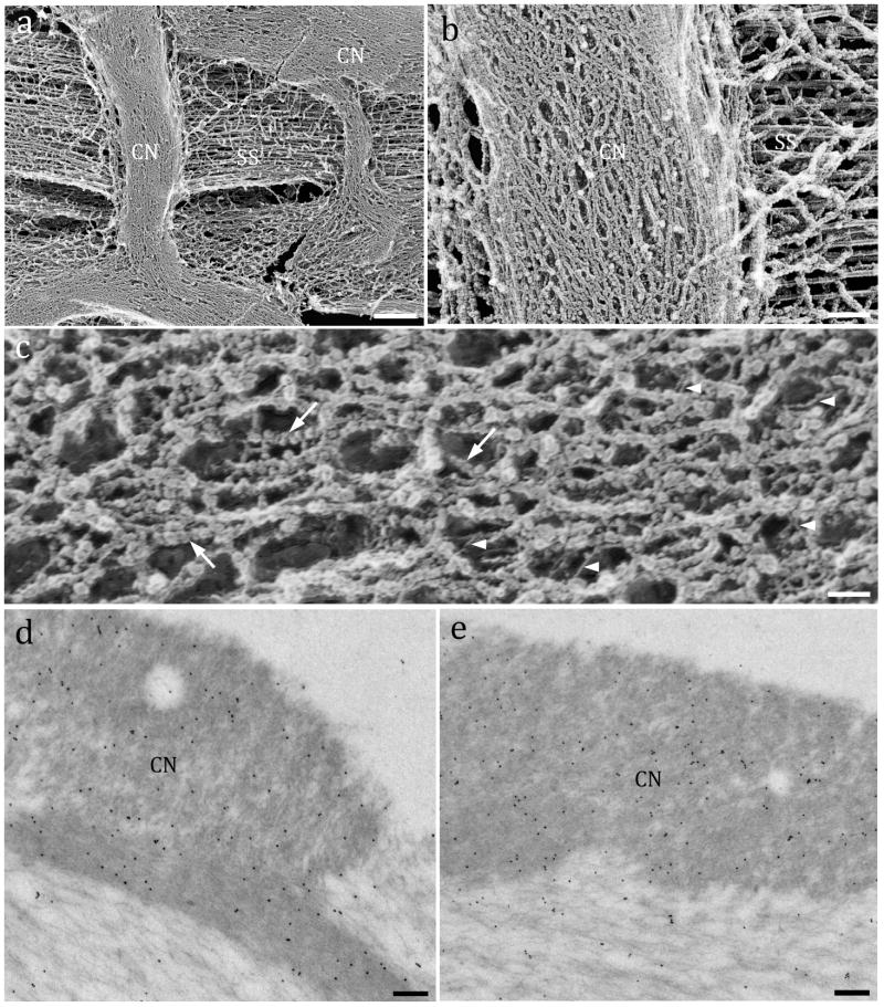
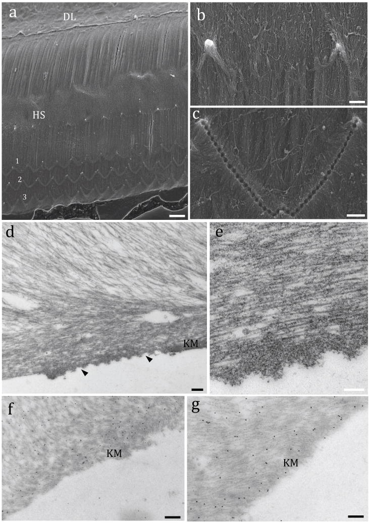
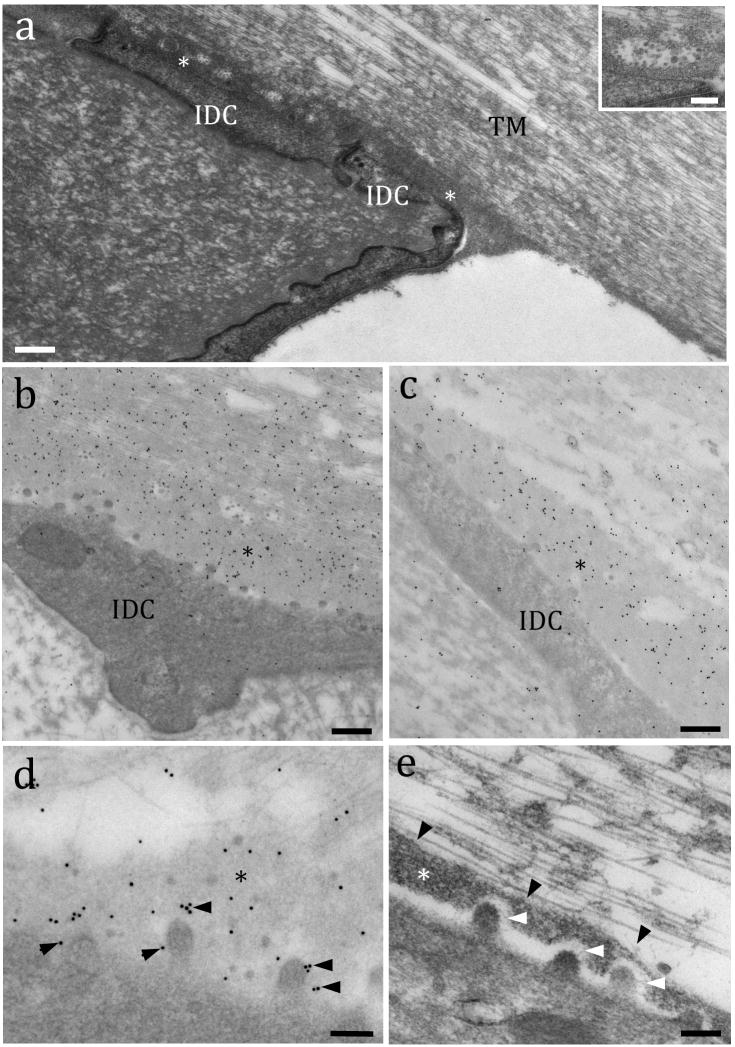
All inner ear organs possess extracellular matrix appendices over the sensory epithelia that are crucial for their proper function. The tectorial membrane (TM) is a gelatinous acellular membrane located above the hearing sensory epithelium and is composed mostly of type II collagen, and α and β tectorins. TM molecules self-assemble in the endolymph fluid environment, interacting medially with the spiral limbus and distally with the outer hair cell stereocilia. Here, we used immunogold labeling in freeze-substituted mouse cochleae to assess the fine localization of both tectorins in distinct TM regions. We observed that the TM adheres to the spiral limbus through a dense thin matrix enriched in α- and β-tectorin, both likely bound to the membranes of interdental cells. Freeze-etching images revealed that type II collagen fibrils were crosslinked by short thin filaments (4±1.5nm, width), resembling another collagen type protein, or chains of globular elements (15±3.2nm, diameter). Gold-particles for both tectorins also localized adjacent to the type II collagen fibrils, suggesting that these globules might be composed essentially of α- and β-tectorins. Finally, the presence of gold-particles at the TM lower side suggests that the outer hair cell stereocilia membrane has a molecular partner to tectorins, probably stereocilin, allowing the physical connection between the TM and the organ of Corti.

Keywords: Electron microscopy; Extracellular matrix; Immunogold-labeling; Tectorial membrane; Tectorins.


Conflict of interest statement

We declare no conflict of interest.

Figures

Figure 1
Figure 1
Tectorial membrane (TM) overview. Fig. 1a: SEM showing color-coded anatomical regions of an adult mouse (Myo15a−/−) TM attached to the spiral limbus (SL) and over the sensory epithelium (SE): blue= limbal region; yellow= cover net; green= striated sheet; red: marginal band; purple: Kimura’s membrane. Fig. 1b: SEM of an adult rat showing the TM upper side cover net (CN) composed of superficial sets of collagen bundles obliquely crossed (arrow), ending on the edge forming the marginal band (MB). Fig. 1c: Side view of an adult mouse TM underlying the sensory epithelium showing the edge of the marginal band (MB) and the collagen bundles of the covernet (CN). Fig. 1d: TEM of a similar region depicted in 1c showing a densely packed marginal band which continues its upside with the cover net bundles and downside by the Kimura’s membrane (KM). Scale bars: a = 15 μm; b and c = 10 μm; d = 2 μm.
Figure 2
Figure 2
Fine structure of the TM striated sheet: Fig. 2a: TEM of a rat TM showing the parallel-arranged type II collagen fibrils interwoven by thin filaments (arrowheads). Inset: TEM of an individual collagen fibril with its typical banding pattern. Fig. 2b: High-resolution freeze-etching image of a guinea pig TM formed by collagen fibrils crosslinked by numerous filamentous materials. Fig. 2c: Detail image of collagen fibrils crosslinked by thin filaments (arrowheads). Fig. 2d: High magnification showing collagen fibrils connected by globular-arranged filaments. Scale bars: a = 100 nm; Inset, b, c and d = 50 nm
Figure 3
Figure 3
Localization of tectorins in the TM striated sheet. Fig. 3a: ImmunoEM for mouse α-tectorin reveals triangular (white arrowheads) and linear patterns (black arrowheads) of gold-particles. Fig. 3b: Higher magnification image of the α-tectorin gold labeling. Arrowheads indicate rows of gold particles. Fig. 3c: ImmunoEM for mouse β-tectorin positive labeling along the entire TM structure but concentrated in more amorphous regions, forming straight (black arrowheads) and triangular shapes (white arrowheads). Fig. 3d: Detail of the β-tectorin labeling in the striated sheet. Fig. 3e: TEM image of a rat TM showing the parallel collagen fibers and amorphous intermediate materials similar to such regions labeled for tectorins. Fig. 3f: Deep-etch image of guinea pig TM showing the collagen fibrils crosslinked by thin filaments (arrowheads) and chains of globules that resemble the structures that are labeled with antibodies to tectorins (arrows). Scale bars: a = 150 nm; b = 100 nm; c = 150 nm; d = 120 nm; e and f = 100 nm.
Figure 4
Figure 4
Fine structure and immunogold labeling of the covernet. Fig. 4a: Freeze-etching of a guinea pig TM showing covernet bundles (CN) above the striated sheet fibers (SS). Fig. 4b: Higher magnification image of the covernet bundle orthogonally crossing parallel collagen fibers (SS) underneath. Fig. 4c: Detail of the covernet fine structure showing branched filaments made of globular units (arrows) and interconnecting thin fibrils (arrowheads). Immunogold TEM for α-tectorin (Fig. 4d) and β-tectorin (Fig. 4e) showing an intense labeling in the covernet (electrondense upper regions) and lesser labeling at the area underneath. Scale bars: a = 2 μm; b = 150 nm; c = 25 nm; d and e = 100 nm.
Figure 5
Figure 5
Ultrastructure and immunogold labeling of the TM lower side. Fig 5a: Low magnification SEM image of the lower side of an adult mouse showing part of the dense lamina (DL), the superficial layer of parallel-arranged collagen bundles with the Hensen’s stripe (HS) and three rows of OHC stereocilia imprints (1, 2, 3). Fig. 5b: Detail of the Hensen’ stripe showing the ridge made by two IHCs. Fig. 5c: Detail of the W shaped imprints generated by the insertion of the tips of the tallest row of OHC stereocilia. Fig. 5d: TEM of the electron dense area of Kimura’s membrane (KM) with stereocilia imprints (arrowheads). Fig. 5e: A detail of the wavelike format of the amorphous material present at the OHC imprints below the fibrillar region of the striated sheet. Immunogold labeling indicated the presence of Tecta (Fig. 5f) and Tectb (Fig. 5g) in the amorphous material of Kimura’s membrane (KM) and upper regions. Scale bars: a = 5 μm; b = 8 μm; b = 6 μm; d = 150 nm; e, f and g = 100 nm.
Figure 6
Figure 6
The TM adhesion to the spiral limbus is mediated by a dense lamina rich in tectorins. Fig 6a: TEM from an adult rat showing the interface between the TM made of collagen parallel fibers and the spiral limbus (at its edge region) formed by a dense lamina layer of an extracellular matrix (*). IDC = interdental cells. Inset: Detail of an exocytotic vesicle found within the dense lamina filled with globular materials. Fig. 6b: Immunogold labeling shows a high concentration of α-tectorin (Fig. 6b) and β-tectorin (Fig. 6c) in the basal lamina (*). Fig 6d: Detail of the α-tectorin labeling close to the microvilli (arrowheads) in the dense lamina (*). Fig. 6e: High magnification TEM showing a detail of the molecular interaction (white arrowheads) between a microvillus and the dense lamina (*), and between the lamina and TM collagen fibrils (black arrowheads). Scale bars: a, b and c = 150 nm; d and e = 100 nm; inset in a = 50 nm.

References

    1. Deans MR, Peterson JM, Wong GW. Mammalian Otolin: a multimeric glycoprotein specific to the inner ear that interacts with otoconial matrix protein Otoconin-90 and Cerebellin-1. PlosOne. 2010;5(9):e12765. - PMC - PubMed
    1. Fichard A, Kleman JP, Ruggiero F. Another look at collagenV and XI molecules. Matrix Biol. 1995;14:515–31. - PubMed
    1. Gavara N, Manoussaki D, Chadwick RS. Auditory mechanics of the tectorial membrane and the cochlear spiral. Curr Opin Otolaryngol Head Neck Surg. 2011;19(5):382–7. - PMC - PubMed
    1. Ghaffari R, Aranyosi AJ, Freeman DM. Longitudinally propagating traveling waves of the mammalian tectorial membrane. Proc Natl Acad Sci USA. 2007;104(42):16510–5. - PMC - PubMed
    1. Ghaffari R, Aranyosi AJ, Richardson GP, Freeman DM. Tectorial membrane travelling waves underlie abnormal hearing in Tectb mutant mice. Nat Commun. 2010;1:96–99. - PMC - PubMed

Publication types

MeSH terms

LinkOut - more resources