

Review
doi: 10.1038/nrn.2015.22.
Rediscovering area CA2: unique properties and functions
Affiliations
- PMID: 26806628
- PMCID: PMC4856153
- DOI: 10.1038/nrn.2015.22
Item in Clipboard
Review
Rediscovering area CA2: unique properties and functions
Nat Rev Neurosci.
2016 Feb.
Abstract
Hippocampal area CA2 has several features that distinguish it from CA1 and CA3, including a unique gene expression profile, failure to display long-term potentiation and relative resistance to cell death. A recent increase in interest in the CA2 region, combined with the development of new methods to define and manipulate its neurons, has led to some exciting new discoveries on the properties of CA2 neurons and their role in behaviour. Here, we review these findings and call attention to the idea that the definition of area CA2 ought to be revised in light of gene expression data.
Conflict of interest statement
The authors declare no competing interests.
Figures
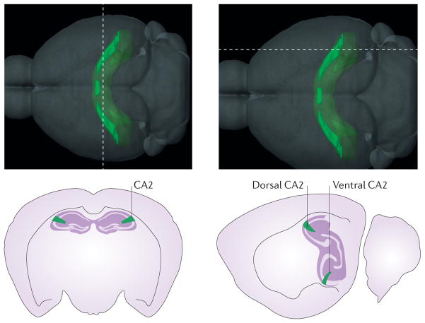
The top panel illustrates the location and gross structure of the mouse CA2 region, as defined by the Allen Institute for Brain Science on the basis of the differential expression of almost 50 genes. Thedashed lines indicate the positions of the slices that are represented in the bottom panels. Interestingly, most of these ‘CA2-enriched’ genes are not expressed in the ventral hippocampus of the mouse (lower right panel), even though other atlases have classically included CA2 in most of the ventral third of the hippocampus. Images in the top panel are reproduced with permission from © 2015 Allen Institute for Brain Science. Allen Mouse Brain Atlas [Internet]. Available from: http://mouse.brain-map.org .
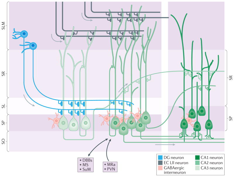
The CA2 region (medium green) is located between the CA3 region (light green) and the CA1 region (dark green) and is an integral part of the hippocampal circuitry. In rodents, the primary afferents contacting CA2 pyramidal neurons arise from three primary sources: dentate gyrus (DG) granule cells (shown in blue) that target the stratum lucidum (SL), CA3 neurons that target the stratum radiatum (SR), and medial and lateral entorhinal cortex layer II (EC LII) neurons (shown in dark grey) that target the stratum lacunosum-moleculare (SLM),,. This part of the perforant path was found to dip far into the area normally considered to be SR in monkeys and humans. The main projection target of CA2 pyramidal neurons is ‘deep’ calbindin 1- immunonegative CA1 neurons with dendrites in stratum oriens (SO),; CA2 neurons target the CA1 SR to a lesser extent. Extrahippocampal inputs to CA2 (indicated by black arrows) include those from vasopressinergic neurons in the paraventricular nucleus (PVN) of the hypothalamus, and from the median raphe (MRa), and reciprocal connections with the supramammillary nucleus (SuM),,, medial septum (MS) and diagonal bands of Broca (DBBs). Many different types of inhibitory interneurons are found in area CA2 and form synapses on CA2 pyramidal neurons. The density of reelin- and parvalbumin-immunopositive interneurons (shown in pink) is several-fold higher in area CA2 stratum pyramidale (SP) than in CA1 SP and CA3 SP.
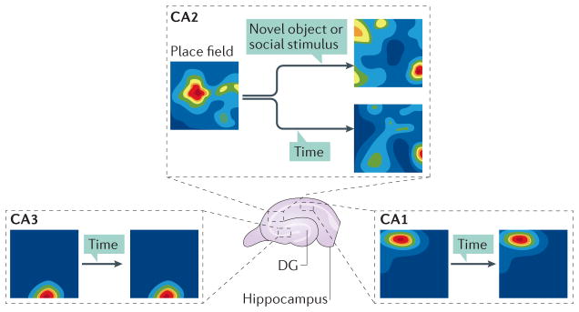
Neuronal recordings in awake behaving rats to characterize spatial firing in CA2(REF. 85) revealed that CA2 neurons fired in place fields, indicated by the warm colours. CA2 place fields differed from those in CA1 and proximal CA3 in that there were more of them and that they were larger. Not illustrated is that several properties of place fields in distal CA3 (closest to CA2; CA3a) closely resembled those in CA2 (REFS 87,88). Notably, place fields in CA2 changed with the passage of time (hours to days) and could shift significantly in response to social stimulation and novel objects (that is, global remapping). These results together suggest that CA2 is less sensitive to novel global contextual cues than to novel local cues and social stimuli. DG, dentate gyrus.
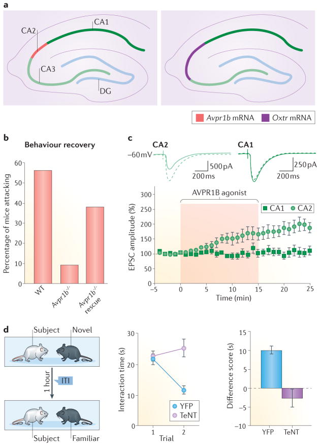
a | The receptors for ‘social’ neuropeptides vasopressin and oxytocin (arginine vasopressin receptor 1B (AVPR1B) and oxytocin receptor (OXTR)) are highly expressed in CA2 pyramidal neurons. BothAvpr1b−/− and Oxtr−/− mice have deficits in social recognition memory and social aggression,. b | Virus-mediated re-expression of Avpr1b in the dorsal CA2 of Avpr1b−/− mice rescued social aggression behaviours. c | An AVPR1B agonist, as well as oxytocin (not illustrated), induces a slowly developing synaptic potentiation in CA2, but not in CA1, neurons. d | Silencing of CA2 pyramidal neuron output in mice impairs social recognition memory, but not sociability. Control animals spent less time investigating familiar mice than novel mice, but the animals with tetanus toxin (TeNT) expressed in CA2 were unable to differentiate between novel and familiar mice. EPSC, excitatory postsynaptic current; ITI, inter-trial interval; WT, wild type; YFP, yellow fluorescent protein. Partsb and c are from REF. , Nature Publishing Group. Part d is from REF. , Nature Publishing Group.
References
-
- van Strien NM, Cappaert NLM, Witter MP. The anatomy of memory: an interactive overview of the parahippocampal–hippocampal network. Nat Rev Neurosci. 2009;10:272–282. - PubMed
-
- Geva-Sagiv M, Las L, Yovel Y, Ulanovsky N. Spatial cognition in bats and rats: from sensory acquisition to multiscale maps and navigation. Nat Rev Neurosci. 2015;16:94–108. - PubMed
Publication types
MeSH terms
Grants and funding
LinkOut - more resources
Full Text Sources
Other Literature Sources
Miscellaneous
