

The reliability of ultrasonography in developmental dysplasia of the hip: How reliable is it in different hands?
- PMID: 26806967
- PMCID: PMC4705726
- DOI: 10.4103/0019-5413.168753
The reliability of ultrasonography in developmental dysplasia of the hip: How reliable is it in different hands?
Abstract
Background: Developmental dysplasia of the hip (DDH) is the most common skeletal dysplasia. Two principal methods used in early diagnosis of DDH are clinical examination and ultrasonographic investigation. Dogruel et al. found a low specificity of clinical examination in patients with DDH. Additionally, Kamath et al. stated that ultrasonography performed by a radiologist in routine clinical practice is more reliable than physical examination performed by the average clinician. In clinical practice, the application and assessment of hip ultrasonography are completed by a single person. This assessment determines the followup of the patient. Thus, hip ultrasonography performed on the same person by different individuals under the same conditions will yield a more accurate assessment of the reliability of ultrasonographic assessment of DDH. Although inter-observer reliability was high in many previous studies of ultrasound image evaluation, reliability rates vary among studies of the application of ultrasonography.
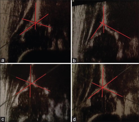
Materials and methods: Inter-examiner reliability of hip ultrasonography was analyzed among four investigators who separately evaluated 100 hips (50 infants). The obtained bone structure angles α, cartilage structure angles β, and distribution of hip types were compared among the investigators. All infants were brought to the hospital for a healthy child followup examination, according to the country's health policy. Babies between 0 and 6 months were included in the study. Babies with any neuromuscular disorders, neural tube defects or any type of genetic anomalies were excluded from the study. The study was explained to the families of all infants and written informed consent was obtained.
Results: There was a significant difference in the hip type determined by the investigators with respect to α and β angles (P < 0.01, P < 0.01, P = 0.002). The average alpha measurements of the first orthopedist, second orthopedist, first radiologist, and second radiologist were 67.38 ± 6.24, 65.60 ± 5.84, 65.44 ± 4.59, and 62.59 ± 4.50, respectively. The average beta measurements of the first orthopedist, second orthopedist, first radiologist, and second radiologist were 53.85 ± 8.86, 50.74 ± 7.80, 44.77 ± 6.30, and 44.39 ± 5.81, respectively. Agreement among the results obtained by the clinicians was investigated in dual comparisons. The relative agreement according to the alpha angle ranged from 3.6% to 44.5%, and the relative concordance according to the beta angle ranged from 0.9% to 45.3%. Agreement regarding hip typing was determined to range from 19.1% to 42.6%.
Conclusion: Sonographic evaluation of the hip appears to vary depending on the investigator.
Keywords: Developmental dysplasia of hip; Hip dysplasia; congenital; hip dislocation; hip ultrasonography; reliability; ultrasonic diagnosis; ultrasonography.
Conflict of interest statement
Figures
References
-
- Kamath S, Mehdi A, Wilson N, Duncan R. The lack of evidence of the effect of selective ultrasound screening on the incidence of late developmental dysplasia of the hip in the Greater Glasgow Region. J Pediatr Orthop B. 2007;16:189–91. - PubMed
-
- Graf R. The diagnosis of congenital hip-joint dislocation by the ultrasonic Combound treatment. Arch Orthop Trauma Surg. 1980;97:117–33. - PubMed
-
- Graf R. Classification of hip joint dysplasia by means of sonography. Arch Orthop Trauma Surg. 1984;102:248–55. - PubMed
LinkOut - more resources
Full Text Sources
Other Literature Sources
Research Materials