

Dspp mutations disrupt mineralization homeostasis during odontoblast differentiation
- PMID: 26807185
- PMCID: PMC4697717
Dspp mutations disrupt mineralization homeostasis during odontoblast differentiation
Abstract
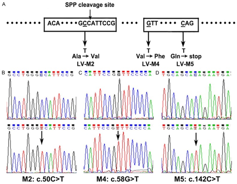
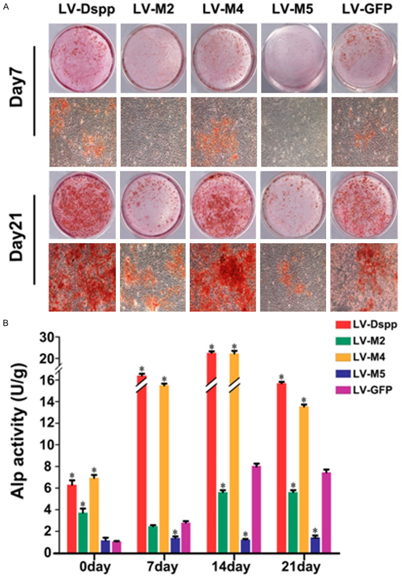
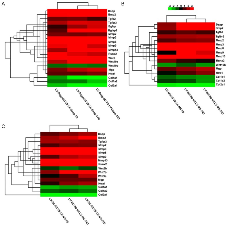
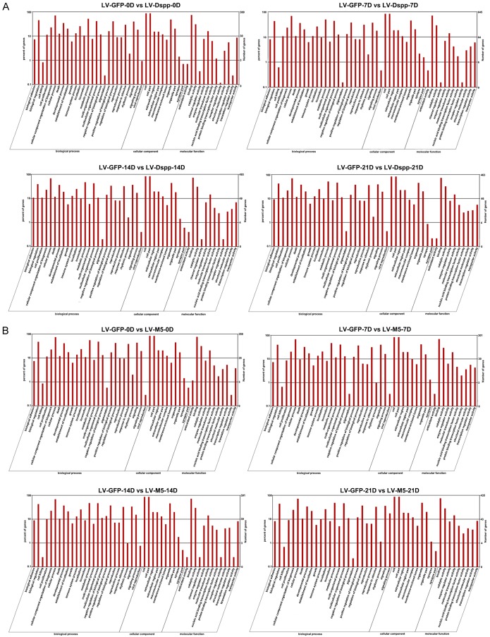
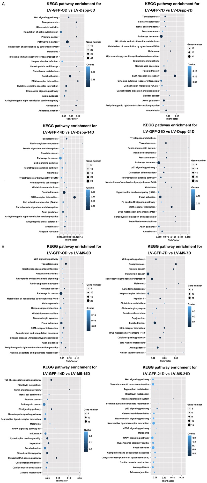
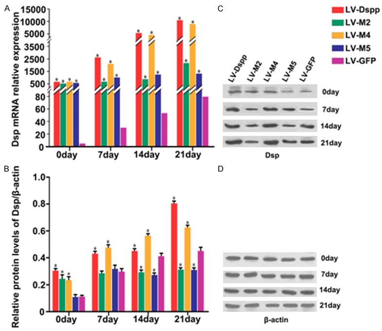
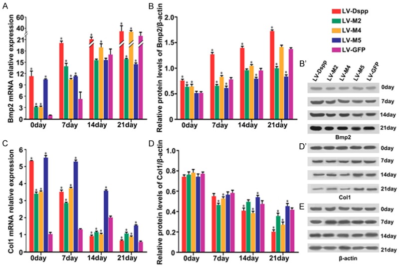
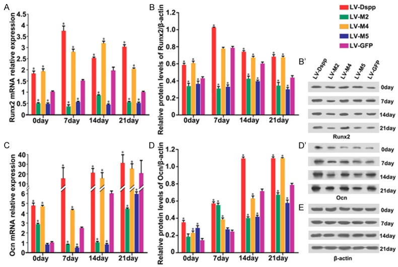
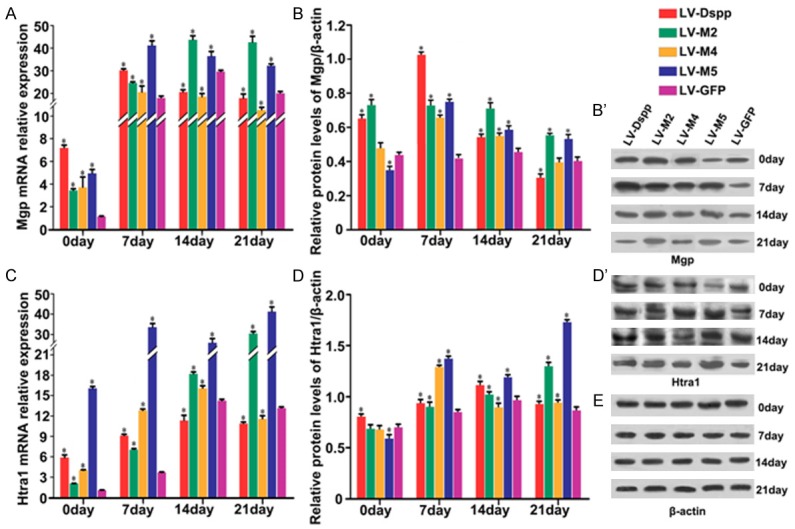
The main pathological feature in isolated hereditary dentin disorders is the abnormality of dentin mineralization. Dentin sialophosphoprotein (DSPP) gene is the only identified causative gene for the disorders. The present study aims to explore the molecular association between Dspp mutations and the disrupted mineralization homeostasis during odontoblast differentiation. We generated lentivirus constructs with the mouse full-length wild type Dspp cDNA and 3 Dspp mutants and transfected them into mouse odontoblast-lineage cells (OLCs) which were then performed 21-day mineralization inducing differentiation. The formation of mineralized nodules was obviously fewer in mutants. Digital Gene Expression (DGE) showed that Dspp mutation affected the OLC differentiation in a degree. Further examination validated that Dspp (LV-Dspp) overexpressing OLCs possessed the ability to strictly orchestrate framework for mineralization inductors like Bmp2, Col1 and Runx2, and proliferative markers for mineralization like Alp and Ocn, as well as mineral homeostasis feedback regulators Mgp and Htra1. However, the missense mutation in Dspp signal peptide region (LV-M2) and the nonsense mutation (LV-M5) broke this orchestration. The results suggested that the mutant Dspp disrupt the dynamic homeostasis of mineralization during OLC differentiation. We are the first to use full-length mouse Dspp gene expression system to explore the mineralization mechanism by which inductors and inhibitors adjust each other during odontoblast differentiation. Our findings shed new light on association between Dspp and the dynamic homeostasis of mineralization inductors and inhibitors, and indicate the disruption of mineralization homeostasis might be a crucial reason for Dspp mutations resulting in dentin disorders.
Keywords: Dentin sialophosphoprotein; dentin; mineralization; mutation; odontoblast-lineage cells (OLCs).
Figures
References
-
- MacDougall M, Simmons D, Luan X, Nydegger J, Feng J, Gu TT. Dentin phosphoprotein and dentin sialoprotein are cleavage products expressed from a single transcript coded by a gene on human chromosome 4. Dentin phosphoprotein DNA sequence determination. J Biol Chem. 1997;272:835–842. - PubMed
-
- Butler WT, Bhown M, Brunn JC, D’Souza RN, Farach-Carson MC, Happonen RP, Schrohenloher RE, Seyer JM, Somerman MJ, Foster RA, et al. Isolation, characterization and immunolocalization of a 53-kDal dentin sialoprotein (DSP) Matrix. 1992;12:343–351. - PubMed
-
- George A, Bannon L, Sabsay B, Dillon JW, Malone J, Veis A, Jenkins NA, Gilbert DJ, Copeland NG. The carboxyl-terminal domain of phosphophoryn contains unique extended triplet amino acid repeat sequences forming ordered carboxyl-phosphate interaction ridges that may be essential in the biomineralization process. J Biol Chem. 1996;271:32869–32873. - PubMed
-
- George A, Hao J. Role of phosphophoryn in dentin mineralization. Cells Tissues Organs. 2005;181:232–240. - PubMed
-
- Wu L, Zhu F, Wu Y, Lin Y, Nie X, Jing W, Qiao J, Liu L, Tang W, Zheng X, Tian W. Dentin sialophosphoprotein-promoted mineralization and expression of odontogenic genes in adipose-derived stromal cells. Cells Tissues Organs. 2008;187:103–112. - PubMed
LinkOut - more resources
Full Text Sources
Miscellaneous