

Mechanical and cellular processes driving cervical myelopathy
- PMID: 26807352
- PMCID: PMC4716567
- DOI: 10.5312/wjo.v7.i1.20
Mechanical and cellular processes driving cervical myelopathy
Abstract
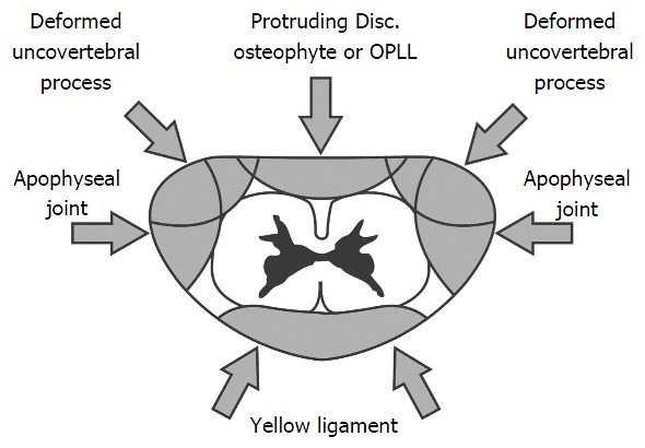
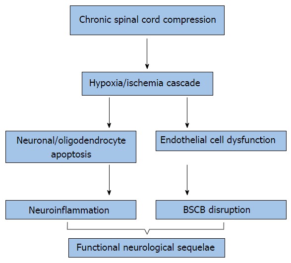
Cervical myelopathy is a well-described clinical syndrome that may evolve from a combination of etiological mechanisms. It is traditionally classified by cervical spinal cord and/or nerve root compression which varies in severity and number of levels involved. The vast array of clinical manifestations of cervical myelopathy cannot fully be explained by the simple concept that a narrowed spinal canal causes compression of the cord, local tissue ischemia, injury and neurological impairment. Despite advances in surgical technology and treatment innovations, there are limited neuro-protective treatments for cervical myelopathy, which reflects an incomplete understanding of the pathophysiological processes involved in this disease. The aim of this review is to provide a comprehensive overview of the key pathophysiological processes at play in the development of cervical myelopathy.
Keywords: Cervical myelopathy; Cervical spine; Neck pain.
Figures

References
-
- Baptiste DC, Fehlings MG. Pathophysiology of cervical myelopathy. Spine J. 2006;6:190S–197S. - PubMed
-
- Yu WR, Liu T, Kiehl TR, Fehlings MG. Human neuropathological and animal model evidence supporting a role for Fas-mediated apoptosis and inflammation in cervical spondylotic myelopathy. Brain. 2011;134:1277–1292. - PubMed
-
- Toledano M, Bartleson JD. Cervical spondylotic myelopathy. Neurol Clin. 2013;31:287–305. - PubMed
-
- Lee J, Satkunendrarajah K, Fehlings MG. Development and characterization of a novel rat model of cervical spondylotic myelopathy: the impact of chronic cord compression on clinical, neuroanatomical, and neurophysiological outcomes. J Neurotrauma. 2012;29:1012–1027. - PubMed
-
- Broomfield SJ, Da Cruz M, Gibson WP. Cochlear implants and magnetic resonance scans: A case report and review. Cochlear Implants Int. 2013;14:51–55. - PubMed
Publication types
LinkOut - more resources
Full Text Sources
Other Literature Sources
