Review
. 2015 Dec;5(6):898-908.
doi: 10.3978/j.issn.2223-4292.2015.09.01.

Keratoameloblastoma or Kerato-odontoameloblastoma: report of its soft tissue recurrence with literature review

Affiliations
Review

Keratoameloblastoma or Kerato-odontoameloblastoma: report of its soft tissue recurrence with literature review

Ravinder S Bedi et al. Quant Imaging Med Surg. 2015 Dec.

Abstract

Keratoameloblastoma (KA) is a rare histological variant of the ameloblastoma with extensive keratin production within the odontogenic islands as well as in the fibrous stroma. Pindborg first reported it in 1970, since then only 18 cases have been reported in the literature. We report a soft tissue recurrence of KA, involving right posterior region of the lower jaw in a 27-year-old female.

Keywords: Ameloblastoma; ameloblast; keratin; mandible; odontogenic tumor; recurrence.


Conflict of interest statement

Conflicts of Interest: The authors have no conflicts of interest to declare.

Figures

Figure 1
Figure 1
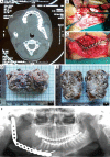
(A) Axial view CT scan showing soft tissue mass on the right side (Arrow marked); (B) tumor mass being exposed; (C) placement of SS reconstruction plate; (D) gross specimen in toto (entire tumor mass in one piece); (E) cut surface revealed numerous cystic spaces with creamish brown cheesy to solid material; (F) post-operative orthopantomograph showing placement of SS reconstruction plate with condylar implant.
Figure 2
Figure 2
(A,B) Imprint cytological smear showing numerous oval to slightly elongated hyperchromatic nucleus with sparse cytoplasm (odontogenic origin) arranged solitary or in aggregates; (C,D) few polygonal cells showing round to oval nucleus with abundant cytoplasm (squamous cell) (Arrow marked). Background stroma consists of flakes of keratin-like material within the sea of RBCs [(A) PAP, ×40; (B-D) PAP, ×100].
Figure 3
Figure 3
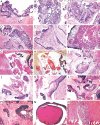
(A) Photomicrograph exhibiting proliferating odontogenic epithelium lining the cystic lumen (H&E, ×40); (B) odontogenic epithelium proliferating in plexiform pattern of odontogenic epithelium (H&E, ×100); (C) papillary projections from odontogenic epithelium (H&E, ×100); (D) papillary proliferation of odontogenic epithelium within a collagenous connective tissue stoma (H&E, ×40); (E) extensive squamous metaplasia with in odontogenic islands lined by tall columnar ameloblast-like cells (H&E, ×100); (F) squamous metaplasia with keratin pearl formation (H&E, ×100); (G) “keratin filled cystic spaces” with keratin production in the stroma (H&E, ×100); (H) “keratin filled cystic spaces” with keratin production in the stroma (Krebergs Stain, ×100); (I) “curvilinear ribbons” of odontogenic epithelium within collagenized stroma, which is extruding a “lamellar stack of keratin” into the stroma without foreign body response (H&E, ×100); (J) “pacinian-like” stack of keratin (H&E, ×100); (K) parakeratin packed elongated epithelial follicles showing lamellar arrangement of keratin forming “hair-like structures” (H&E, ×100); (L) formation of dentinoid-like material adjacent to odontogenic epithelium (H&E, ×100); (M) dentinoid-like material (H&E, ×1,000); (N) dentinoid-like material with tubular structures (H&E, ×1,000); (O) granuloma with cholesterol cleft formation (H&E, ×100).
Figure 4
Figure 4
Distribution of gender (%) (18 published cases of KA). KA, keratoameloblastoma.
Figure 5
Figure 5
Jaw involvement (%) (18 published cases of KA). KA, keratoameloblastoma.
Figure 6
Figure 6
Distribution of location (%) (18 published cases of KA). KA, keratoameloblastoma.
Figure 7
Figure 7
Distribution of KA sub-types (%) (18 published cases of KA). KA, keratoameloblastoma.
Figure 8
Figure 8
Recurrences rate (%) (18 published cases of KA). KA, keratoameloblastoma.

References

    1. Pindborg JJ, Weinmann JP. Squamous cell metaplasia with calcification in ameloblastomas. Acta Pathol Microbiol Scand 1958;44:247-52. - PubMed
    1. Whitt JC, Dunlap CL, Sheets JL, Thompson ML. Keratoameloblastoma: a tumor sui generis or a chimera? Oral Surg Oral Med Oral Pathol Oral Radiol Endod 2007;104:368-76. - PubMed
    1. Pindborg JJ. Pathology of the dental hard tissues. Philadelphia(PA): W. B. Saunders;1970:381-2.
    1. Altini M, Lurie R, Shear M. A case report of keratoameloblastoma. Int J Oral Surg 1976;5:245-9. - PubMed
    1. Altini M, Slabbert HD, Johnston T. Papilliferous keratoameloblastoma. J Oral Pathol Med 1991;20:46-8. - PubMed