

Uptake of 18F-DCFPyL in Paget's Disease of Bone, an Important Potential Pitfall in Clinical Interpretation of PSMA PET Studies
- PMID: 26807444
- PMCID: PMC4721507
- DOI: 10.18383/j.tom.2015.00169
Uptake of 18F-DCFPyL in Paget's Disease of Bone, an Important Potential Pitfall in Clinical Interpretation of PSMA PET Studies
Abstract
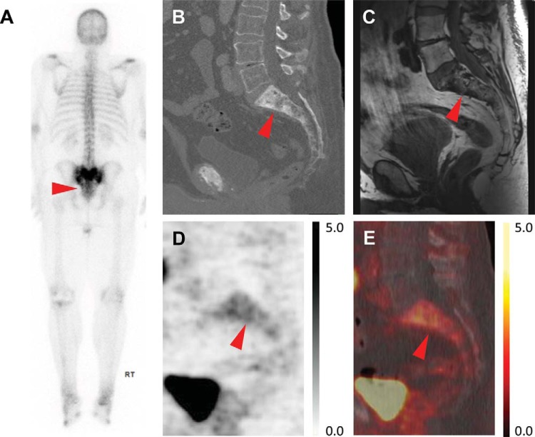
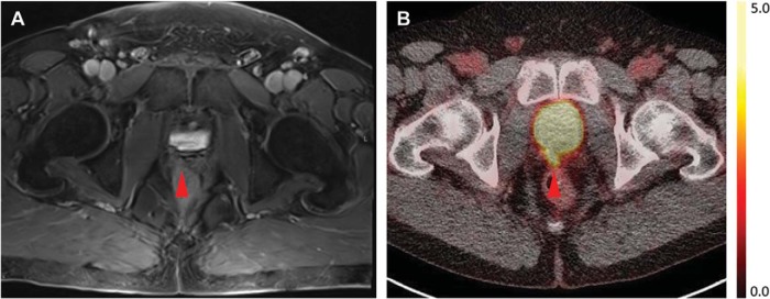
Prostate-specific membrane antigen (PSMA)-targeted PET imaging is an emerging technique for evaluating patients with prostate cancer (PCa) in a variety of clinical contexts. As with any new imaging modality, there are interpretive pitfalls that are beginning to be recognized. In this image report, we describe the findings in a 63-year-old male with biochemically recurrent PCa after radical prostatectomy who was imaged with 18F-DCFPyL, a small molecule inhibitor of PSMA. Diffuse radiotracer uptake was noted throughout the sacrum, corresponding to imaging findings on contrast-enhanced CT, bone scan, and pelvic MRI consistent with Paget's disease of bone. The uptake of 18F-DCFPyL in Paget's disease is most likely due to hyperemia and increased radiotracer delivery. In light of the overlap in patients affected by PCa and Paget's, it is important for nuclear medicine physicians and radiologists interpreting PSMA PET/CT scans to be aware of the potential for this diagnostic pitfall. Correlation to findings on conventional imaging such as diagnostic CT and bone scan can help confirm the diagnosis.
Keywords: DCFPyL; PET/CT; PSMA; Prostate cancer.
Conflict of interest statement
Conflicts of Interest: M.G.P. is a coinventor of a US patent covering [18F]DCFPyL and as such is entitled to a portion of any licensing fees and royalties generated by this technology. This arrangement has been reviewed and approved by the Johns Hopkins University in accordance with its conflict of interest policies.
Figures
References
-
- Cho SY, Gage KL, Mease RC, Senthamizhchelvan S, Holt DP, Jeffrey-Kwanisai A, Endres CJ, Dannals RF, Sgouros G, Lodge M, Eisenberger MA, Rodriguez R, Carducci MA, Rojas C, Slusher BS, Kozikowski AP, Pomper MG. Biodistribution, tumor detection, and radiation dosimetry of 18F-DCFBC, a low-molecular-weight inhibitor of prostate-specific membrane antigen, in patients with metastatic prostate cancer. J Nucl Med. 2012;53:1883–1891. - PMC - PubMed
-
- Afshar-Oromieh A, Malcher A, Eder M, Eisenhut M, Linhart G, Hadaschik BA, Holland-Letz T, Giesel FL, Kratochwil C, Haufe S, Haberkorn U, Zechmann CM. PET imaging with a [68Ga]gallium-labelled PSMA ligand for the diagnosis of prostate cancer: biodistribution in humans and first evaluation of tumour lesions. Eur J Nucl Med Mol Imaging. 2013;40:486–495. - PubMed
-
- Szabo Z, Mena E, Rowe SP, Plyku D, Nidal R, Eisenberger MA, Antonarakis ES, Fan H, Dannals RF, Chen Y, Mease RC, Vranesic M, Bhatnagar A, Sgouros G, Cho SY, Pomper MG. Initial evaluation of [18F]DCFPyL for prostate-specific membrane antigen (PSMA)-targeted PET imaging of prostate cancer. Mol Imaging Biol. 2015;17:565–574. - PMC - PubMed
-
- Wright GL Jr., Grob BM, Haley C, Grossman K, Newhall K, Petrylak D, Troyer J, Konchuba A, Schellhammer PF, Moriarty R. Upregulation of prostate-specific membrane antigen after androgen-deprivation therapy. Urology. 1996;48:326–334. - PubMed
-
- Sweat SD, Pacelli A, Murphy GP, Bostwick DG. Prostate-specific membrane antigen expression is greatest in prostate adenocarcinoma and lymph node metastases. Urology. 1998;52:637–640. - PubMed
Grants and funding
LinkOut - more resources
Full Text Sources
Other Literature Sources
Research Materials
Miscellaneous