. 2016 Mar:92:1-9.
doi: 10.1016/j.yjmcc.2016.01.019. Epub 2016 Jan 22.

Increased fibroblast chymase production mediates procollagen autophagic digestion in volume overload

Affiliations

Increased fibroblast chymase production mediates procollagen autophagic digestion in volume overload

Lianwu Fu et al. J Mol Cell Cardiol. 2016 Mar.

Abstract

Background: Previous work has identified mast cells as the major source of chymase largely associated with a profibrotic phenotype. We recently reported increased fibroblast autophagic procollagen degradation in a rat model of pure volume overload (VO). Here we demonstrate a connection between increased fibroblast chymase production and autophagic digestion of procollagen in the pure VO of aortocaval fistula (ACF) in the rat.

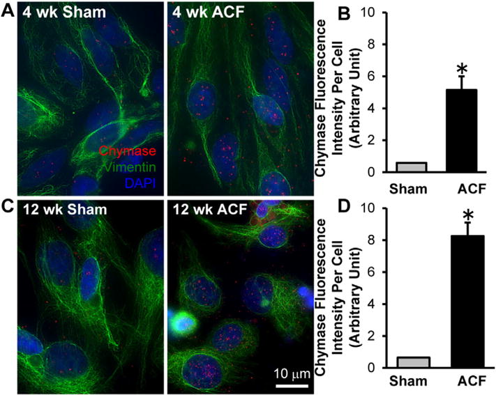
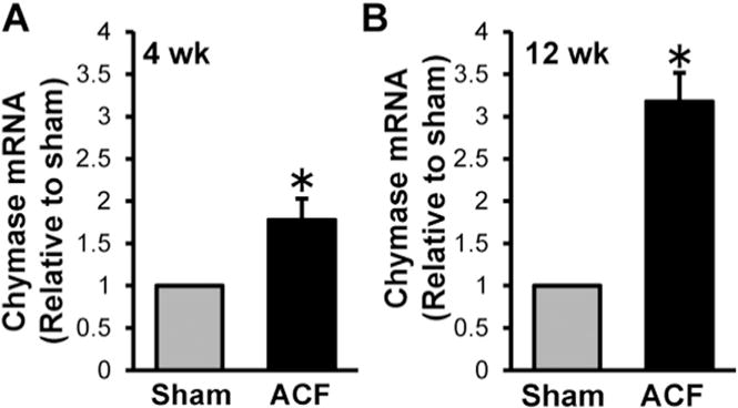
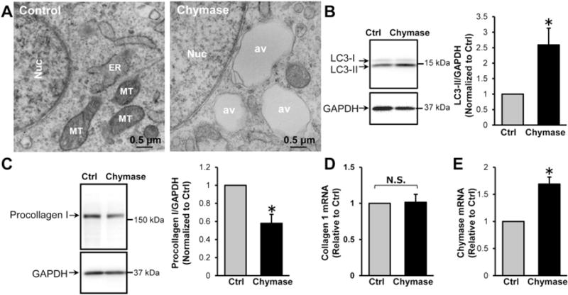
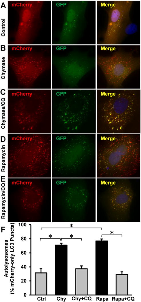
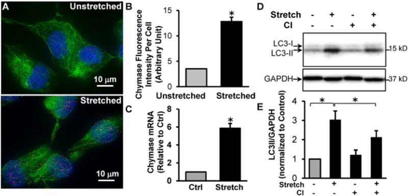
Methods and results: Isolated LV fibroblasts taken from 4 and 12week ACF Sprague-Dawley rats have significant increases in chymase mRNA and chymase activity. Increased intracellular chymase protein is documented by immunocytochemistry in the ACF fibroblasts compared to cells obtained from age-matched sham rats. To implicate VO as a stimulus for chymase production, we show that isolated adult rat LV fibroblasts subjected to 24h of 20% cyclical stretch induces chymase mRNA and protein production. Exogenous chymase treatment of control isolated adult cardiac fibroblasts demonstrates that chymase is internalized through a dynamin-dependent mechanism. Chymase treatment leads to an increased formation of autophagic vacuoles, LC3-II production, autophagic flux, resulting in increased procollagen degradation. Chymase inhibitor treatment reduces cyclical stretch-induced autophagy in isolated cardiac fibroblasts, demonstrating chymase's role in autophagy induction.

Conclusion: In a pure VO model, chymase produced in adult cardiac fibroblasts leads to autophagic degradation of newly synthesized intracellular procollagen I, suggesting a new role of chymase in extracellular matrix degradation.

Keywords: Autophagy; Cardiac fibroblast; Chymase; Intracellular procollagen; Volume overload.


Figures

Fig. 1
Fig. 1
Chymase protein is increased in cardiac fibroblasts isolated from 4 and 12 week ACF rats compared to age-matched shams as shown by Immunocytochemistry. Cardiac fibroblasts were isolated from 4 week (A) and 12 week (C) sham and ACF rats followed by indirect fluorescence microcopy using chymase (red) and vimentin (green) antibodies. DAPI: blue. Quantitation of chymase fluorescence intensity in at least 40 cells demonstrates a marked increase in chymase protein in fibroblasts isolated from 4 week (B) and 12 week (D) ACF rats compared to the age-matched shams.
Fig. 2
Fig. 2
Chymase activity is increased in cardiac fibroblasts isolated from 4 and 12 week ACF rats compared to age-matched shams. Cardiac fibroblasts were isolated from 4 week (A) and 12 week (B) sham and ACF rats were analyzed for chymase activity as described in the Experimental methods section. (A, B) Representative HPLC chromatograms showing chymase activity based on 125I-Ang-II formation from the hydrolysis of 125I-Ang-(1–12). The increased chymase activity is blocked when chymostatin is presented in the reaction mixture (C). (D) Bar graph demonstrates that chymase activity is increased in cardiac fibroblast cells isolated from 4 and 12 week ACF rats compared to age-matched shams. n = 4 each. There is also a significant increase in chymase activity in 12 week ACF fibroblasts vs. 4 week ACF fibroblasts. *P < 0.05.
Fig. 3
Fig. 3
Chymase mRNA is increased in cardiac fibroblasts isolated from 4 and 12 week ACF rats compared to age-matched shams. Cardiac fibroblasts were isolated from 4 week (A) and 12 week (B) sham and ACF rats were analyzed for chymase mRNA by real-time PCR as described in the Experimental methods section. There is a marked increase of chymase mRNA in cardiac fibroblasts isolated from ACF rats. n = 6 each. *P < 0.05.
Fig. 4
Fig. 4
Exogenous chymase is internalized to adult cardiac fibroblasts through dynamin-mediated endocytosis. (A), Adult rat cardiac fibroblast cells were incubated with recombinant human chymase (2.5 μg/ml) for 2 h followed by ICC analysis with anti-chymase (red) and anti-vimentin (green) antibodies. There is marked chymase entry into the cytoplasm and nuclei of the fibroblasts (left panel) that is prevented by pretreatment with dynasore, a dynamin inhibitor (80 μM, right panel). (B), Control experiments reveal that dynasore treatment prevents transferrin uptake by fibroblasts. Rat cardiac fibroblasts were incubated with 5 μg/ml transferrin-Alexa 594 (red) for 2 h followed by fluorescence microscopy. There is marked entry of transferrin into the fibroblast cytoplasm (left panel) that is prevented by pretreatment with dynasore (right panel). n = 3. DAPI: blue.
Fig. 5
Fig. 5
Chymase treatment induces autophagy and procollagen degradation in cardiac fibroblasts. (A), Adult rat cardiac fibroblasts were treated with chymase (2.5 μg/ml) for 2 h and then processed for TEM analyses. There is a marked increase of autophagic vacuoles. av: autophagic vacuoles; Nuc: nucleus; ER: rough endoplasmic reticulum; MT: mitochondria. (B), The expression of an autophagic marker, LC3-II, was analyzed by immunoblotting. Chymase treatment increases LC3-II expression, n = 4. (C), Chymase treatment results in a 50% reduction of procollagen I protein as measured by immunoblotting with no change in collagen I mRNA as shown by RT-PCR (D), n = 4, N.S.: not significant. (E) Chymase treatment increases chymase mRNA as shown by RT-PCR. n = 4, *P < 0.05 vs. control.
Fig. 6
Fig. 6
Chymase induces autophagic flux in adult cardiac fibroblasts. Adult cardiac fibroblast cells grown on 4-well chamber slides were transfected with the plasmid expressing mCherry-GFP-LC3 as described in the Experimental methods. 24 h after transfection, the cells were treated for 2 h with (A) mock control, (B) 2.5 μg/ml recombinant chymase (Chy), (C) Chymase and 100 μM chloroquine (CQ), (D) 5 μM Rapamycin (Rapa), or (E) 5 μM Rapamycin and CQ. The formation of autophagosomes and autolysosomes during autophagy process was analyzed by fluorescence microscopy. (F), Quantitation of red-only puncta in at least 40 transfected cells from each group demonstrates an increased autophagic flux in cardiac fibroblasts treated with chymase or rapamycin. *P < 0.05.
Fig. 7
Fig. 7
Mechanical stretch of adult rat cardiac fibroblasts induces chymase production and autophagy that is inhibited by a chymase inhibitor. Adult rat LV fibroblasts cultured in Flexcell plates were subjected to 20% cyclic stretch (1 Hz) for 24 h. (A), Representative images of ICC shows an increased chymase protein in stretched fibroblasts. Chymase: red; Vimentin: green; Blue: DAPI. (B), Quantitation of fluorescence intensity of at least 40 cells demonstrates a 3.6-fold increase of chymase protein upon stretch. (C), Quantitative RT-PCR shows a ~ 6 fold increase of chymase mRNA after stretch. (D), Adult cardiac fibroblasts were pretreated with 100 μM chymase inhibitor (TEI-F00806) and subjected to cyclic stretch for 24 h. Western blot demonstrates that stretch-induced LC3-II production is attenuated by chymase inhibitor. (E), Bar graph shows the results of 4 experiments. *P < 0.05.

Comment in

References

    1. Wei CC, Chen Y, Powell LC, Zheng J, Shi K, Bradley WE, et al. Cardiac kallikreinkinin system is upregulated in chronic volume overload and mediates an inflammatory induced collagen loss. PLoS ONE. 2012;7:e40110. - PMC - PubMed
    1. Ryan TD, Rothstein EC, Aban I, Tallaj JA, Husain A, Lucchesi PA, et al. Left ventricular eccentric remodeling and matrix loss are mediated by bradykinin and precede cardiomyocyte elongation in rats with volume overload. J Am Coll Cardiol. 2007;49:811–821. - PubMed
    1. Chen YW, Pat B, Gladden JD, Zheng J, Powell P, Wei CC, et al. Dynamic molecular and histopathological changes in the extracellular matrix and inflammation in the transition to heart failure in isolated volume overload. Am J Physiol Heart Circ Physiol. 2011;300:H2251–H2260. - PMC - PubMed
    1. Dell’Italia LJ, Meng QC, Balcells E, Wei CC, Palmer R, Hageman GR, et al. Compartmentalization of angiotensin II generation in the dog heart. Evidence for independent mechanisms in intravascular and interstitial spaces. J Clin Invest. 1997;100:253–258. - PMC - PubMed
    1. Dell’Italia LJ, Husain A. Dissecting the role of chymase in angiotensin II formation and heart and blood vessel diseases. Curr Opin Cardiol. 2002;17:374–379. - PubMed

Publication types

MeSH terms