. 2016 Mar;12(3):188-93.
doi: 10.1038/nchembio.2008. Epub 2016 Jan 25.

A two-state activation mechanism controls the histone methyltransferase Suv39h1

Affiliations

A two-state activation mechanism controls the histone methyltransferase Suv39h1

Manuel M Müller et al. Nat Chem Biol. 2016 Mar.

Abstract

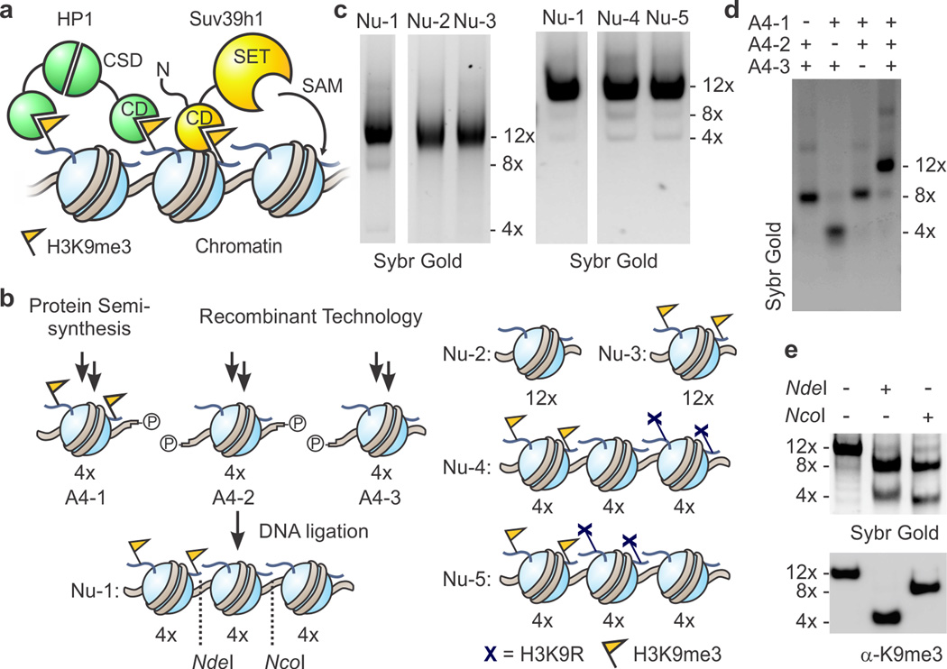
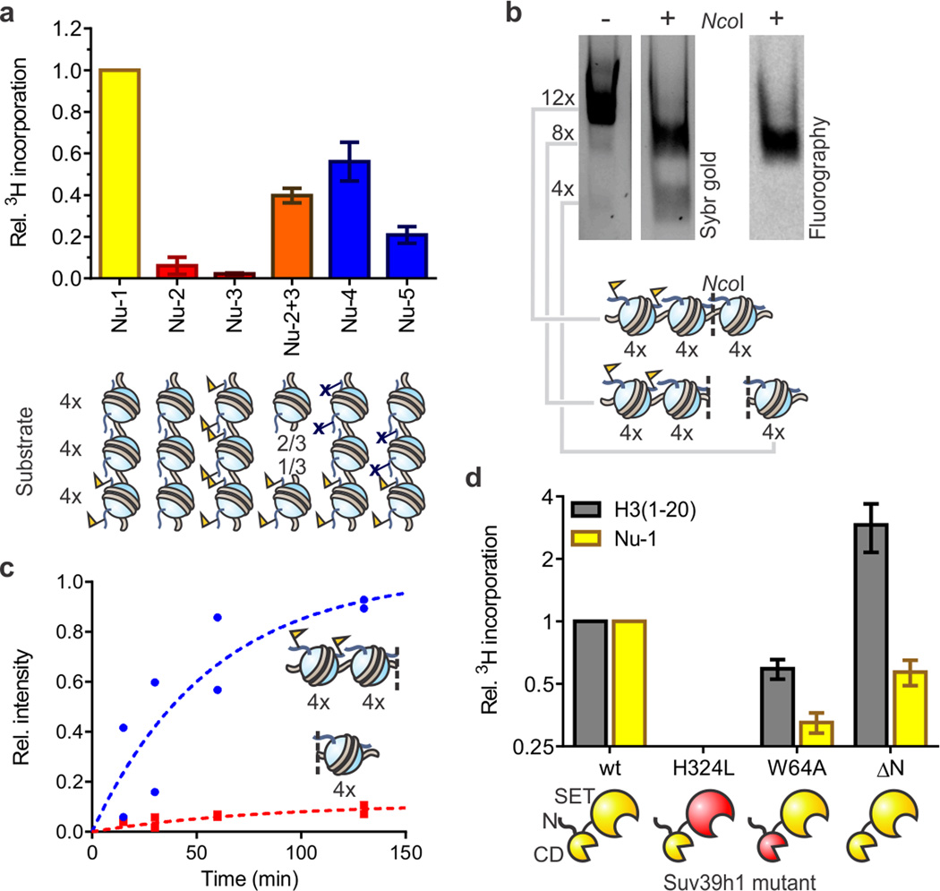
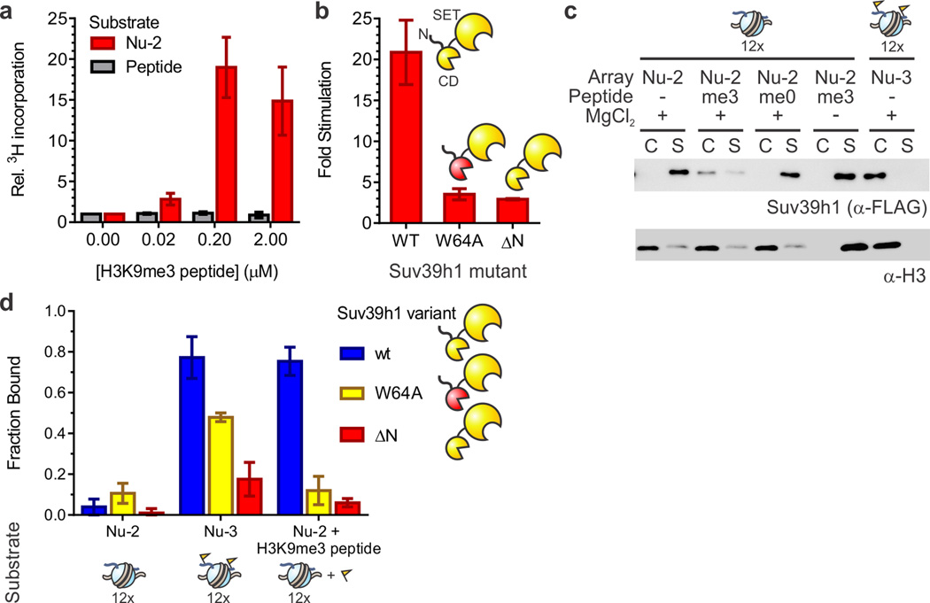
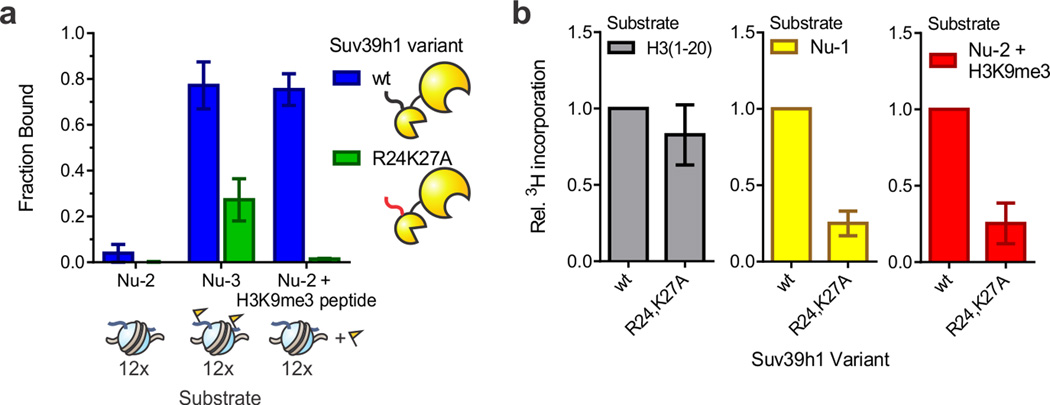
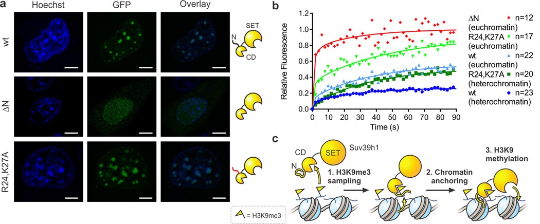
Specialized chromatin domains contribute to nuclear organization and regulation of gene expression. Gene-poor regions are di- and trimethylated at lysine 9 of histone H3 (H3K9me2 and H3K9me3) by the histone methyltransferase Suv39h1. This enzyme harnesses a positive feedback loop to spread H3K9me2 and H3K9me3 over extended heterochromatic regions. However, little is known about how feedback loops operate on complex biopolymers such as chromatin, in part because of the difficulty in obtaining suitable substrates. Here we describe the synthesis of multidomain 'designer chromatin' templates and their application to dissecting the regulation of human Suv39h1. We uncovered a two-step activation switch where H3K9me3 recognition and subsequent anchoring of the enzyme to chromatin allosterically promotes methylation activity and confirmed that this mechanism contributes to chromatin recognition in cells. We propose that this mechanism serves as a paradigm in chromatin biochemistry, as it enables highly dynamic sampling of chromatin state combined with targeted modification of desired genomic regions.


Figures

Figure 1
Figure 1. Heterotypic designer chromatin substrates
(a) Simplified model for heterochromatin spreading. H3K9me3-modified (yellow FLAGs) nucleosomes serve to recruit the scaffolding protein HP1 and the histone methyltransferase (HMT) Suv39h1 through their chromodomains (CDs). The latter catalyzes the S-adenosylmethionine (SAM)-dependent methylation of H3K9 via its SET domain, thus providing a positive feedback loop. HP1 forms a dimer through interactions of its chromo-shadow domain (CSD). (b) Schematic for the synthesis of heterotypic designer nucleosome arrays. NdeI and NcoI sites for restriction analysis are indicated by dotted lines. See supporting information for more detail. Cartoon representations of control arrays are shown on the right. (c) Purity of designer chromatin assessed by native gel electrophoresis. For additional data, see Supplementary Fig. 3. (d) Ligations of purified 4-mer arrays proceed selectively as indicated by native gel electrophoresis of crude products from ligation reactions of designated array fragments. (e) PTM-containing ‘priming’ domain remains intact through designer chromatin synthesis. Ligated arrays (Nu-1) were digested and the resulting fragments analyzed by native gel electrophoresis and western blotting for H3K9me3. See supplementary Figure 3c for more detail.
Figure 2
Figure 2. Reconstitution of Suv39h1-dependent heterochromatin spreading in vitro
(a) Substrate preference of Suv39h1-catalyzed spreading. Arrays Nu-2 and Nu-3 represent homotypic templates (red) carrying either H3K9me0 or H3K9me3, respectively. Inter-fiber spreading is evaluated with a 2:1 mixture of arrays Nu-2 and Nu-3 (orange) to match the stoichiometry of histones in array Nu-1 (yellow). Arrays Nu-4 and Nu-5 (blue) contain H3K9R mutations to block spreading. HMT activity was measured using 3H-SAM as the co-factor in the presence of 5 mM MgCl2. Scintillation counts are normalized to the values determined with heterotypic array Nu-1, without additional normalization for number of substrate sites. Error bars, s.e.m. (n = 3). (b) Suv39h1 preferentially methylates nucleosomes adjacent to preinstalled H3K9me3 marks. Arrays were methylated with 3H-SAM in the presence of 0.5 mM MgCl2 and subsequently digested with NcoI, separated by native gel electrophoresis and analyzed by Sybr gold staining (left) and fluorography (right). See Supplementary Fig. 8 for more details. (c) Time course of Suv39h1-dependent spreading of the H3K9me3 mark. Individual values, normalized to the sum of the values for the 4mer and 8mer obtained at 130 min, from two independent measurements are shown. (d) HMT activity of Suv39h1 variants with an H3 peptide encompassing residues 1–20 (gray) or array Nu-1 (yellow). Measurements are normalized to the values obtained with wild-type Suv39h1 and the respective substrate, and plotted on a log2 scale. Error bars, s.e.m. (n = 3).
Figure 3
Figure 3. Trans-activation of Suv39h1
(a) Allosteric activation of Suv39h1 by H3K9me3 peptides occurs only on chromatin substrates. 3H-SAM-based HMT assays were performed on either unmodified peptide (gray) or array Nu-2 (red) substrates in the presence of increasing amounts of H3K9me3 peptide added in trans. In each case, 3H-me incorporation was normalized to the value obtained in the absence of H3K9me3 peptide. Error bars, s.e.m. (n = 4). (b) Suv39h1-activation requires an intact chromodomain and N-terminus. Stimulation of HMT activity is defined as the ratio of scintillation counts obtained on unmodified chromatin Nu-2 in the presence of 0.2 µM H3K9me3 peptide vs. its absence. Error bars, s.e.m. (n = 3). (c) The H3K9me3 mark promotes chromatin binding in cis and in trans. Western blot of a binding assay of wild-type Suv39h1 with array Nu-2 or Nu-3. Where appropriate, H3 peptides are added at a concentration of 0.2 µM. A control reaction in the absence of MgCl2 is included (lanes 7 and 8). See Supplementary Fig. 14 for full image of the membrane. (d) Chromatin binding is promoted by the H3K9me3 mark and requires an intact chromodomain and N-terminus. Bound fractions are determined by densitometry of Suv39h1 western blots (α-FLAG) bands corresponding to the re-dissolved pellet (chromatin associated) and supernatant (unbound) of binding reactions. Error bars, s.e.m. (n = 3).
Figure 4
Figure 4. The N-terminus of Suv39h1 contributes to chromatin binding in vitro
(a) Residues R24 and K27 of Suv39h1 contribute to chromatin binding. Bound fractions are determined by densitometry of Suv39h1 western blots (α-FLAG) bands corresponding to the re-dissolved pellet (chromatin associated) and supernatant (unbound) of co-precipitation reactions. Error bars, s.e.m. (n = 3). (b) The R24,K27A mutant displays reduced activity on chromatin substrates. HMT measurements with H3 peptides (left), array Nu-1 (middle), or array Nu-2 in the presence of 0.2 µM H3K9me3 peptide (right) were normalized to the values obtained for wt Suv39h1. Error bars, s.e.m. (n = 5).
Figure 5
Figure 5. The Suv39h1 N-terminus contributes to chromatin binding in vivo
(a) Fluorescence microscopy images of indicated Suv39h1-GFP fusion proteins in NIH-3T3 cells. Cells were stained with Hoechst 33342 dye to visualize nuclei and heterochromatin foci. Scale bar = 5 µM. (b) FRAP analysis of Suv39h1-GFP fusion proteins in NIH-3T3 cells. FRAP profiles were recorded upon bleaching heterochromatin foci or a circular spot in the low intensity region of nuclei (euchromatin). For visualization purposes, the average of 12–23 individual measurements were fit to the coupled reaction-diffusion model of Sprague et al. (ref ). (c) Model for a two-state activation of Suv39h1 methyltransferase activity. Freely diffusing Suv39h1 exhibits low HMT activity (left). Chromodomain (CD)-dependent recognition of nucleosomal H3K9me3 and subsequent anchoring to chromatin involving the N-terminus of Suv39h1 (center) enhances HMT activity in the vicinity of the stimulating mark (right).

Similar articles

Cited by

References

    1. Kornberg RD. Structure of chromatin. Annu Rev Biochem. 1977;46:931–954. - PubMed
    1. Woodcock CL, Ghosh RP. Chromatin higher-order structure and dynamics. Cold Spring Harb Perspect Biol. 2010;2:1–25. - PMC - PubMed
    1. Rea S, et al. Regulation of chromatin structure by site-specific histone H3 methyltransferases. Nature. 2000;406:593–599. - PubMed
    1. Nakayama J, Rice JC, Strahl BD, Allis CD, Grewal SI. Role of histone H3 lysine 9 methylation in epigenetic control of heterochromatin assembly. Science. 2001;292:110–113. - PubMed
    1. Grewal SI, Moazed D. Heterochromatin and epigenetic control of gene expression. Science. 2003;301:798–802. - PubMed

ONLINE METHODS REFERENCES

    1. Blanco-Canosa JB, Dawson PE. An Efficient Fmoc-SPPS Approach for the Generation of Thioester Peptide Precursors for Use in Native Chemical Ligation. Angew Chem Int Ed. 2008;47:6851–6855. - PMC - PubMed
    1. Wan Q, Danishefsky SJ. Free-radical-based, specific desulfurization of cysteine: a powerful advance in the synthesis of polypeptides and glycopolypeptides. Angew Chem Int Ed. 2007;46:9248–9252. - PubMed
    1. Dyer PN, Edayathumangalam RS, White CL, Bao Y, Chakravarthy S, Muthurajan UM, Luger K. Reconstitution of nucleosome core particles from recombinant histones and DNA. Methods Enzymol. 2004;375:23–44. - PubMed
    1. Dorigo B, Schalch T, Bystricky K, Richmond TJ. Chromatin fiber folding: Requirement for the histone H4 N-terminal tail. J Mol Biol. 2003;327:85–96. - PubMed
    1. Fierz B, Chatterjee C, McGinty RK, Bar-Dagan M, Raleigh DP, Muir TW. Histone H2B ubiquitylation disrupts local and higher-order chromatin compaction. Nat Chem Biol. 2011;7:113–119. - PMC - PubMed

Publication types