. 2016 Jan 25;11(1):e0146658.
doi: 10.1371/journal.pone.0146658. eCollection 2016.

Real-Time Investigation of Tuberculosis Transmission: Developing the Respiratory Aerosol Sampling Chamber (RASC)

Affiliations

Real-Time Investigation of Tuberculosis Transmission: Developing the Respiratory Aerosol Sampling Chamber (RASC)

Robin Wood et al. PLoS One. .

Abstract

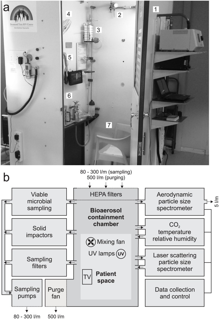
Knowledge of the airborne nature of respiratory disease transmission owes much to the pioneering experiments of Wells and Riley over half a century ago. However, the mechanical, physiological, and immunopathological processes which drive the production of infectious aerosols by a diseased host remain poorly understood. Similarly, very little is known about the specific physiological, metabolic and morphological adaptations which enable pathogens such as Mycobacterium tuberculosis (Mtb) to exit the infected host, survive exposure to the external environment during airborne carriage, and adopt a form that is able to enter the respiratory tract of a new host, avoiding innate immune and physical defenses to establish a nascent infection. As a first step towards addressing these fundamental knowledge gaps which are central to any efforts to interrupt disease transmission, we developed and characterized a small personal clean room comprising an array of sampling devices which enable isolation and representative sampling of airborne particles and organic matter from tuberculosis (TB) patients. The complete unit, termed the Respiratory Aerosol Sampling Chamber (RASC), is instrumented to provide real-time information about the particulate output of a single patient, and to capture samples via a suite of particulate impingers, impactors and filters. Applying the RASC in a clinical setting, we demonstrate that a combination of molecular and microbiological assays, as well as imaging by fluorescence and scanning electron microscopy, can be applied to investigate the identity, viability, and morphology of isolated aerosolized particles. Importantly, from a preliminary panel of active TB patients, we observed the real-time production of large numbers of airborne particles including Mtb, as confirmed by microbiological culture and polymerase chain reaction (PCR) genotyping. Moreover, direct imaging of captured samples revealed the presence of multiple rod-like Mtb organisms whose physical dimensions suggested the capacity for travel deep into the alveolar spaces of the human lung.


Conflict of interest statement

Competing Interests: WAB and CJC are employed by Zeteo Tech LLC. AJH is employed by RTI International. CER is employed by Aerosol Exposure Dimensions. Belonging to these commercial entities does not alter the authors’ adherence to PLOS ONE policies on sharing data and materials.

Figures

Fig 1
Fig 1. The Respiratory Aerosol Sampling Chamber (RASC).
(A) Photograph of the RASC (with the door open) on site in a community TB clinic (1) aerodynamic particle sizer (2) Filter samplers (3) Andersen impactor (4) Mixing fan (5) CO2, temperature and RH (6) PM10 impactor (7) Chair for participant. (B) Block diagram depicting the fluidic and electronic configuration of the RASC. Thick connecting lines indicate airflow and aerosol paths; thin lines indicate electronic connections. All air leaving the RASC is HEPA filtered.
Fig 2
Fig 2. The in-built APS characterizes the particle size distribution spectrum within the RASC.
(A) Typical background particle spectrum before and after air wash. Note the 10-fold decrease in particle counts across all size ranges following the air wash. Total count from a typical five seconds of sampling. (B) Artificial dry release of fluorescent polystyrene latex (PSL) microspheres. The APS instrument groups all particles with an aerodynamic diameter less than 0.523 μm in the number bin on the far left of each chart. Note that the 1μm release (b) contained approximately ten times more particles in the release. Total count from ten seconds of sampling at the peak of particle concentration. (Inset) corresponding SEM images of the released particles.
Fig 3
Fig 3. The effect of aerosol hydration on particle size distribution.
The graph shows APS measurements of “wet” and “dry” releases of M. smegmatis::gfp. (see text for details).
Fig 4
Fig 4. Isolation and visualization of viable mycobacteria in the RASC.
(A) M. smegmatis::gfp growth on solid 7H10 agar plates from the Six-Stage Viable Andersen Cascade Impactor after wet release of 200 μl diluted culture into the RASC (30 000, 3000 and 300 colony forming units—CFU). The columns indicate the particle sizes captured on each plate across the 6 stages of the impactor, and the rows indicate the estimated total number of CFU passing through the impactor. Each release was repeated three times and the mean and SD for each plate are presented below the typical growth pattern distribution seen in the particle release. In all the releases the sampling was run for 5 minutes at 28 l/min resulting in the potential total capture of 3000, 300 and 30 CFU respectively. (B) SEM (left) and fluorescent microscopy (right) of M. smegmatis::gfp isolated on a PM10 impactor following experimental release.
Fig 5
Fig 5. Particle production as a function of respiration in a clinical TB patient.
CO2 concentration (solid line and left ordinate) and particle counts (dots and right ordinate) in the 1–2.5 μm size range for a TB patient.
Fig 6
Fig 6. Isolation of Mtb from a TB patient.
SEM image of patient sample impacted on the lower plate of the PM10 impactor. The dimensions and morphology of the rod-shaped structure (denoted by *) are consistent with the presence of Mtb bacilli in the untreated TB patient. There is also evidence of multiple “splats” of unknown identity (one example is denoted by **) which might comprise organic matter derived from patient lung or respiratory tract. Note the “halo” structures (dark shadows) surrounding each particle.

References

    1. Riley RL, Mills CC, Nyka W, Weinstock N, Storey PB, Sultan LU, et al. (1959) Aerial dissemination of pulmonary tuberculosis. A two-year study of contagion in a tuberculosis ward. Am J Hygiene 70: 2. - PubMed
    1. Riley RL, Mills CC, O'Grady F, Sultan LU, Wittstadt F, Shivpuri DN (1962) Infectiousness of air from a tuberculosis ward. Ultraviolet irradiation of infected air: comparative infectiousness of different patients. Am Rev Respir Dis 85: 511–525. - PubMed
    1. Escombe AR, Moore DAJ, Gilman RH, Pan W, Navincopa M, Ticona E, et al. (2008) The infectiousness of tuberculosis patients coinfected with HIV. PLoS Med 5: e188 10.1371/journal.pmed.0050188 - DOI - PMC - PubMed
    1. Dharmadhikari AS, Basaraba RJ, Van Der Walt ML, Weyer K, Mphahlele M, Venter K, et al. (2011) Natural infection of guinea pigs exposed to patients with highly drug-resistant tuberculosis. Tuberculosis (Edinb) 91: 329–338. - PMC - PubMed
    1. Issarow CM, Mulder N, Wood R (2015) Modelling the risk of airborne infectious disease using exhaled air. J Theor Biol 372: 100–106. 10.1016/j.jtbi.2015.02.010 - DOI - PubMed

Publication types