

In Vivo Dynamic Deformation of Articular Cartilage in Intact Joints Loaded by Controlled Muscular Contractions
- PMID: 26807930
- PMCID: PMC4726761
- DOI: 10.1371/journal.pone.0147547
In Vivo Dynamic Deformation of Articular Cartilage in Intact Joints Loaded by Controlled Muscular Contractions
Abstract
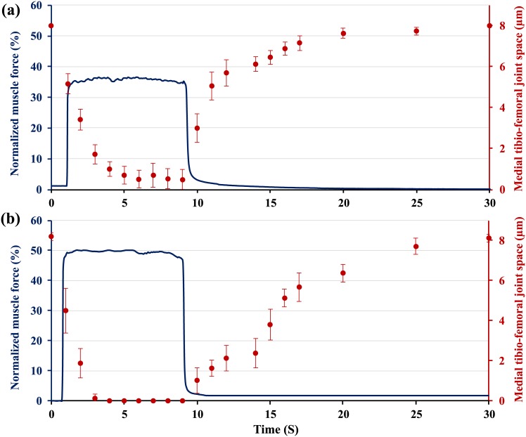
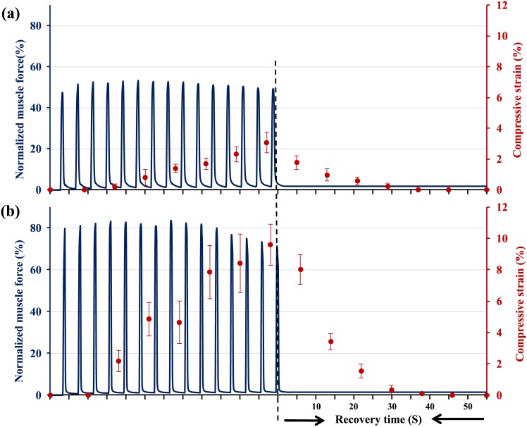
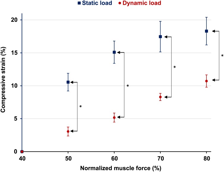
When synovial joints are loaded, the articular cartilage and the cells residing in it deform. Cartilage deformation has been related to structural tissue damage, and cell deformation has been associated with cell signalling and corresponding anabolic and catabolic responses. Despite the acknowledged importance of cartilage and cell deformation, there are no dynamic data on these measures from joints of live animals using muscular load application. Research in this area has typically been done using confined and unconfined loading configurations and indentation testing. These loading conditions can be well controlled and allow for accurate measurements of cartilage and cell deformations, but they have little to do with the contact mechanics occurring in a joint where non-congruent cartilage surfaces with different material and functional properties are pressed against each other by muscular forces. The aim of this study was to measure in vivo, real time articular cartilage deformations for precisely controlled static and dynamic muscular loading conditions in the knees of mice. Fifty and 80% of the maximal knee extensor muscular force (equivalent to approximately 0.4N and 0.6N) produced average peak articular cartilage strains of 10.5±1.0% and 18.3±1.3% (Mean ± SD), respectively, during 8s contractions. A sequence of 15 repeat, isometric muscular contractions (0.5s on, 3.5s off) of 50% and 80% of maximal muscular force produced cartilage strains of 3.0±1.1% and 9.6±1.5% (Mean ± SD) on the femoral condyles of the mouse knee. Cartilage thickness recovery following mechanical compression was highly viscoelastic and took almost 50s following force removal in the static tests.
Conflict of interest statement
Figures

References
-
- Mow VC, Ratcliffe A, Poole AR. Cartilage and Diarthrodial Joints As Paradigms for Hierarchical Materials and Structures. Biomaterials 1992;13(2):67–97. - PubMed
-
- Shrive NG, Frank CB. Articular Cartilage In: Nigg BM, Herzog W, editors. Biomechanics of the Musculo-skeletal System. 2nd Edition ed. London: John Wiley & Sons; 1999. p. 86–106.
-
- Moskowitz RW. Experimental models of osteoarthritis In: Moskowitz RW, Howell DS, Goldberg VM, Mankin HJ, editors. Osteoarthritis, Diagnosis and Management.Philadelphia: W.B.Saunders Company; 1984. p. 213–32.
-
- Guilak F, Ratcliffe A, Mow VC. Chondrocyte deformation and local tissue strain in articular cartilage: a confocal microscopy study. J Orthop Res 1995;13:410–21. - PubMed
-
- Buschmann MD, Hunziker EB, Kim YJ, Grodzinsky AJ. Altered aggrecan synthesis correlates with cell and nucleus structure in statically compressed cartilage. J Cell Sci 1996. February;109 (Pt 2):499–508. - PubMed
Publication types
MeSH terms
Grants and funding
LinkOut - more resources
Full Text Sources
Other Literature Sources
