Comparative Study
. 2016 Jan 25;11(1):e0147198.
doi: 10.1371/journal.pone.0147198. eCollection 2016.

Comparative Study of Injury Models for Studying Muscle Regeneration in Mice

Affiliations
Comparative Study

Comparative Study of Injury Models for Studying Muscle Regeneration in Mice

David Hardy et al. PLoS One. .

Abstract

Background: A longstanding goal in regenerative medicine is to reconstitute functional tissues or organs after injury or disease. Attention has focused on the identification and relative contribution of tissue specific stem cells to the regeneration process. Relatively little is known about how the physiological process is regulated by other tissue constituents. Numerous injury models are used to investigate tissue regeneration, however, these models are often poorly understood. Specifically, for skeletal muscle regeneration several models are reported in the literature, yet the relative impact on muscle physiology and the distinct cells types have not been extensively characterised.

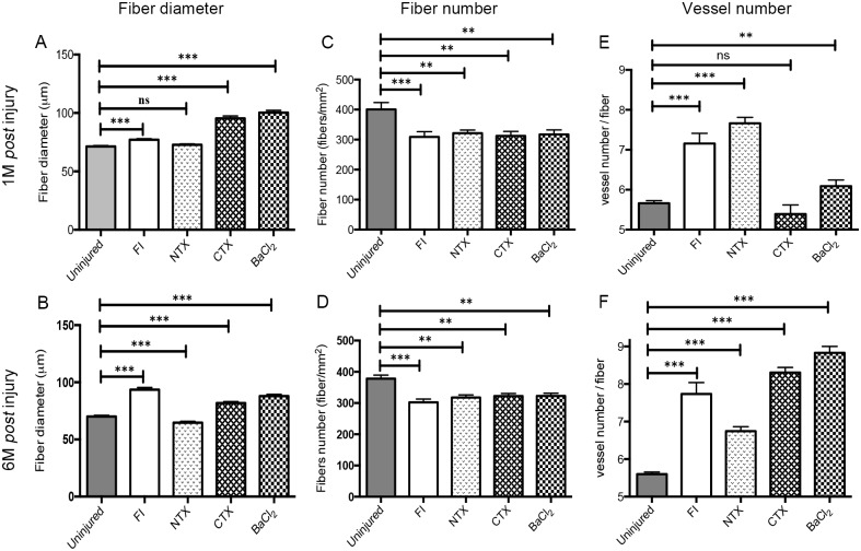
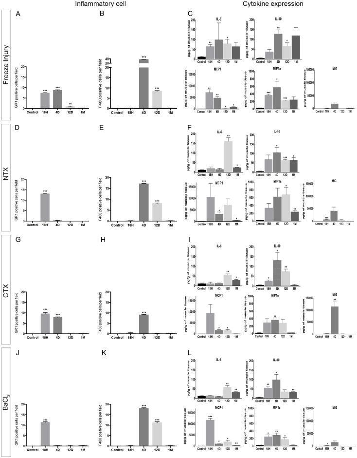
Methods: We have used transgenic Tg:Pax7nGFP and Flk1GFP/+ mouse models to respectively count the number of muscle stem (satellite) cells (SC) and number/shape of vessels by confocal microscopy. We performed histological and immunostainings to assess the differences in the key regeneration steps. Infiltration of immune cells, chemokines and cytokines production was assessed in vivo by Luminex®.

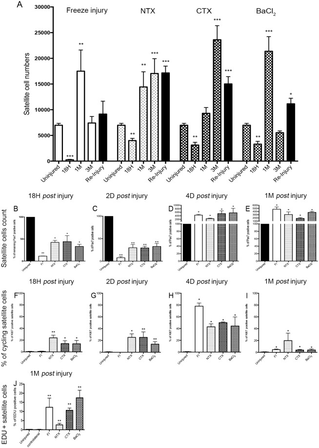
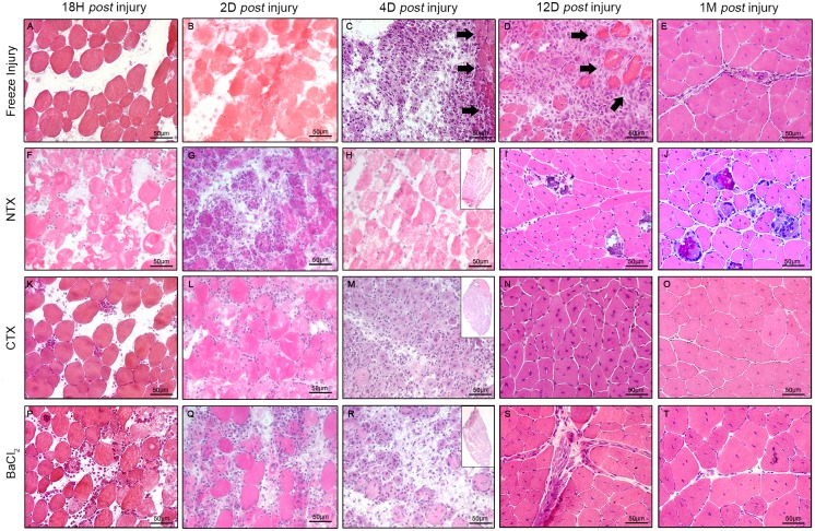
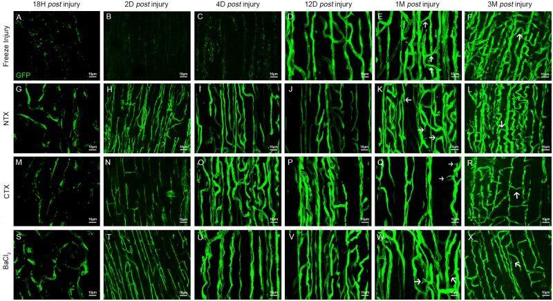
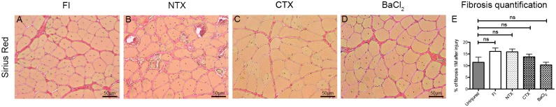
Results: We compared the 4 most commonly used injury models i.e. freeze injury (FI), barium chloride (BaCl2), notexin (NTX) and cardiotoxin (CTX). The FI was the most damaging. In this model, up to 96% of the SCs are destroyed with their surrounding environment (basal lamina and vasculature) leaving a "dead zone" devoid of viable cells. The regeneration process itself is fulfilled in all 4 models with virtually no fibrosis 28 days post-injury, except in the FI model. Inflammatory cells return to basal levels in the CTX, BaCl2 but still significantly high 1-month post-injury in the FI and NTX models. Interestingly the number of SC returned to normal only in the FI, 1-month post-injury, with SCs that are still cycling up to 3-months after the induction of the injury in the other models.

Conclusions: Our studies show that the nature of the injury model should be chosen carefully depending on the experimental design and desired outcome. Although in all models the muscle regenerates completely, the trajectories of the regenerative process vary considerably. Furthermore, we show that histological parameters are not wholly sufficient to declare that regeneration is complete as molecular alterations (e.g. cycling SCs, cytokines) could have a major persistent impact.


Conflict of interest statement

Competing Interests: The authors have declared that no competing interests exist.

Figures

Fig 1
Fig 1. Number of satellite cells and their behaviour in the 4 different injury models.
(A) Number of satellite cells in uninjured, 18h, 1 month and 3 months post-injury; n = 124 animals (ancillary and new data). The figure also displays the number of satellite cells after re-injury (i.e. after two successive lesions carried out 28 days apart; displayed in black histograms) n = 5 animals. (B-E) Percentage of remaining Pax7 positive cells (B) 18h, (C) 2 days, (D) 4 days and (E) 1 month post-injury on TA sections. (F-I) Percentage of cycling Ki67 positive satellite cells (F) 18h, (G) 2 days, (H) 4 days and (I) 1 month after injury. Data are represented as means±s.d. *p < 0.05; **p < 0.01; ***p < 0.001; no star, statistically non significant.
Fig 2
Fig 2. Muscle histology at different time points after injury.
Haematoxylin and eosin staining on cryosections. (A) 18h, (B) 2 days, (C) 4 days (D) 12 days and (E) one month post freeze injury. (F) 18h, (G) 2 days, (H) 4 days (I) 12 days and (J) one month post NTX injury. (K) 18h, (L) 2 days, (M) 4 days (N) 12 days and (O) one month post CTX injury. (P) 18h, (Q) 2 days, (R) 4 days (S) 12 days and (T) one month post BaCl2 injury. Insets represent whole muscle scan (HMR). Scale bar represents 50 μm.
Fig 3
Fig 3. Fibre quantification and vascularization at different time points post injury in the 4 injury models.
(A) Fibre diameters (expressed in μm) 1 month after injury in all 4 injury models (B), 6 months after injury in all 4 injury models (C,D), 1 and 6 months, respectively, in all injury models. (E) Vessel numbers per fibre 1 month after injury in all injury models. (F) Vessel numbers per fibre 6 month after injury for all injury models. Data are represented as means±s.d. *p < 0.05; **p < 0.01; ***p < 0.001; ns, statistically non significant.
Fig 4
Fig 4. Characterization of inflammation after injury in the 4 injury models.
(A,D,G,J) Neutrophil (GR1+ cells) quantifications (expressed as number of cells per ten microscopic fields at 40X) in the FI (A), NTX (D), CTX (G) and BaCl2 (J). (B,E,H,K) Macrophage (F4/80+ cells) quantifications (expressed as number of cells per field) in the FI (B), NTX (E), CTX (H) and BaCl2 (K). Data are represented as means±s.d. *p < 0.05; **p < 0.01; ***p < 0.001; no star, statistically non significant. (C,F,I,L) Luminex (multiplex assay) measuring the levels of cytokines in pg/g of muscle tissue, 18h, 4 days, 12 days and 1 month post-injury in the freeze injury model (C), the NTX (F), the CTX (I), and the BaCl2 (L). Selected cytokines (IL6; IL10; MCP1; MIP1a and MIG) are displayed for each injury model. Data are represented as means±s.d.
Fig 5
Fig 5. Three dimensional analysis of vessels at different time points in all 4 injury models.
Images show blood vessel organisation in 3D after z-stack reconstitutions of scanned sectioned TA from Flk1GFP/+ mouse. (A-F) Vessel organisation in the freeze injury, 18h (A), 2 days (B), 4 days (C), 12 days (D), 1 month (E) and 3 months (F) post injury. (G-L) Vessel organisation in the NTX injury, 18h (G), 2 days (H), 4 days (I), 12 days (J), 1 month (K) and 3 months (L) post injury. (M-R) Vessel organisation in the CTX injury, 18h (M), 2 days (N), 4 days (O), 12 days (P), 1 month (Q) and 3 months (R) post injury. (S-X) Vessel organisation in the BaCl2 injury, 18h (S), 2 days (T), 4 days (U), 12 days (V), 1 month (W) and 3 months (X) post-injury. Arrows pointing anastomoses. Scale bars represents 10 μm.
Fig 6
Fig 6. Characterization of fibrosis after injury.
(A-D) Sirius Red staining (collagen deposits)1 month after injury in all 4 injury models. (E) Percentage of fibrosis per section 1 month after injury compared with non-injured control. No statistically significant differences detected among the 4 models. ns; non significant.

References

    1. Mauro A. Satellite cell of skeletal muscle fibers. J Biophys Biochem Cytol. 1961;9: 493–5. 10.1083/jcb.9.2.493 - DOI - PMC - PubMed
    1. Gayraud-Morel B, Chrétien F, Tajbakhsh S. Skeletal muscle as a paradigm for regenerative biology and medicine. Regen Med. 2009;4: 293–319. 10.2217/17460751.4.2.293 - DOI - PubMed
    1. Tajbakhsh S. Skeletal muscle stem cells in developmental versus regenerative myogenesis. Journal of Internal Medicine. 2009. pp. 372–389. 10.1111/j.1365-2796.2009.02158.x - DOI - PubMed
    1. Zammit PS, Partridge TA, Yablonka-Reuveni Z. The skeletal muscle satellite cell: the stem cell that came in from the cold. J Histochem Cytochem. 2006;54: 1177–91. 10.1369/jhc.6R6995.2006 - DOI - PubMed
    1. Arnold L, Henry A, Poron F, Baba-Amer Y, van Rooijen N, Plonquet A, et al. Inflammatory monocytes recruited after skeletal muscle injury switch into antiinflammatory macrophages to support myogenesis. J Exp Med. 2007;204: 1057–69. 10.1084/jem.20070075 - DOI - PMC - PubMed

Publication types

MeSH terms