. 2016 Apr 15;11(4):1074-81.
doi: 10.1021/acschembio.5b00948. Epub 2016 Feb 5.

HIV-1 Integrase Strand Transfer Inhibitors with Reduced Susceptibility to Drug Resistant Mutant Integrases

Affiliations

HIV-1 Integrase Strand Transfer Inhibitors with Reduced Susceptibility to Drug Resistant Mutant Integrases

Xue Zhi Zhao et al. ACS Chem Biol. .

Abstract

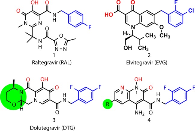
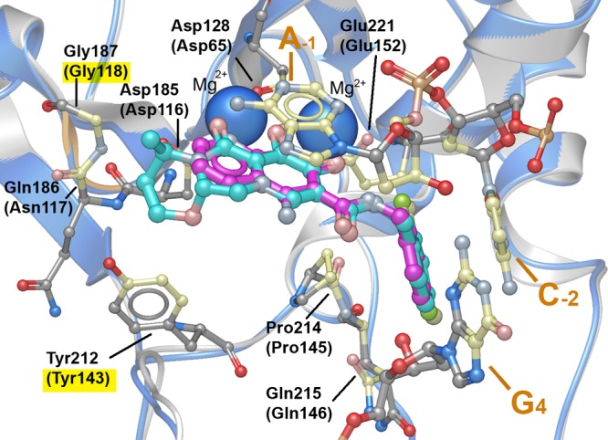
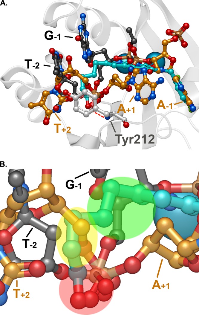
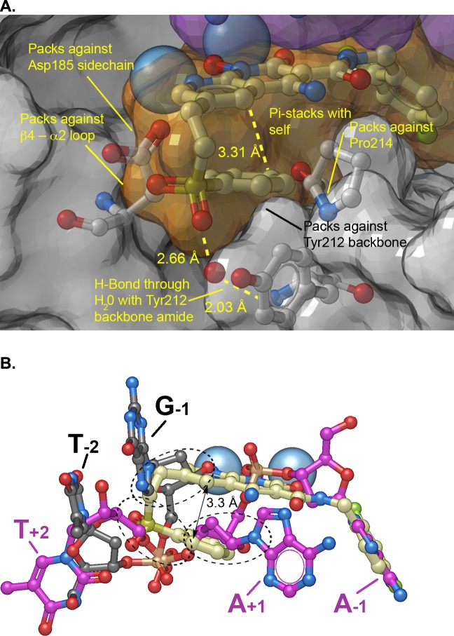
HIV integrase (IN) strand transfer inhibitors (INSTIs) are among the newest anti-AIDS drugs; however, mutant forms of IN can confer resistance. We developed noncytotoxic naphthyridine-containing INSTIs that retain low nanomolar IC50 values against HIV-1 variants harboring all of the major INSTI-resistant mutations. We found by analyzing crystal structures of inhibitors bound to the IN from the prototype foamy virus (PFV) that the most successful inhibitors show striking mimicry of the bound viral DNA prior to 3'-processing and the bound host DNA prior to strand transfer. Using this concept of "bi-substrate mimicry," we developed a new broadly effective inhibitor that not only mimics aspects of both the bound target and viral DNA but also more completely fills the space they would normally occupy. Maximizing shape complementarity and recapitulating structural components encompassing both of the IN DNA substrates could serve as a guiding principle for the development of new INSTIs.


Conflict of interest statement

The authors declare the following competing financial interest(s): Compounds described in the paper are included in pending or filed patent applications.

Figures

Figure 1
Figure 1
Structures of the FDA-approved INSTIs (RAL, 1; EVG, 2; and DTG, 3) and heterobicyclic compounds of the current series (4). Colors highlight key functional features: metal-chelating triad of heteroatoms (red), halobenzyl rings (blue). The terminal ring of DTG and the corresponding 6-substituents described in the present work shown in green shading.
Figure 2
Figure 2
DTG and 4a binding in the active site of the PFV intasome. PFV-bound DTG (cyan with protein ribbon in gray) is shown with Mg2+ ions (solid blue spheres) along with the A-1 and C-2 nucleotides of 3′-processed viral DNA and the G4 nucleotide of the uncleaved complementary strand (PDB code 3S3M). Interacting residues are shown with contacting carbons shown in cream (corresponding IN residues are indicated in parentheses). The protein β4−α2 loop is highlighted in orange. Contact residues associated with RAL resistance (Y143R and G118R) are highlighted in yellow. PFV-bound 4a is superimposed onto the DTG-bound structure with the ligand carbons in magenta and the protein ribbon in blue.
Figure 3
Figure 3
Compound 4c in the active site of the PFV intasome. (A) Superposition of the binding of 4c (blue) with pre-3′-processed DNA (carbons in gold; PDB code: 4E7I(25)) and target DNA (carbons in black; PDB code: 4E7K(25)). The bases of proximal nucleotides of the viral and host DNAs are annotated. The hydrogen bond between the 6- substituent terminal hydroxyl of 4c with the backbone amide nitrogen of Tyr212 is shown. (B) Closeup of panel A highlighting regions of correspondence between the 6-side chain of 4c and the nucleotides of the substrate DNAs: red, phosphoryl oxygens; yellow, T+2 deoxysugar; and green, G–1 deoxysugar. The IN residue Tyr212 is not shown for clarity.
Figure 4
Figure 4
PFV Intasome crystal structure complexed with 4f. (A) Crystal structure oriented to show the complexed Mg2+ ions (blue spheres) and the semitransparent surface of 4f (orange surface with carbons in cream), 3′P DNA (purple surface), and protein (gray surface) with key protein residues shown (carbons in light gray). Important binding interactions are indicated. (B) Bound 4f with superimposed pre-3′-processed DNA (carbons in purple; PDB code: 4E7I(25)) and target DNA (carbons in black; PDB code: 4E7K(25)). Regions where the side chain of 4f overlaps with DNA are indicated by black dashed ovals.

Similar articles

Cited by

References

    1. Pommier Y.; Marchand C. (2012) Interfacial inhibitors: Targeting macromolecular complexes. Nat. Rev. Drug Discovery 11, 25–36. 10.1038/nrd3404. - DOI - PMC - PubMed
    1. Cherepanov P.; Maertens G. N.; Hare S. (2011) Structural insights into the retroviral DNA integration apparatus. Curr. Opin. Struct. Biol. 21, 249–256. 10.1016/j.sbi.2010.12.005. - DOI - PubMed
    1. Di Santo R. (2014) Inhibiting the HIV integration process: Past, present, and the future. J. Med. Chem. 57, 539–566. 10.1021/jm400674a. - DOI - PMC - PubMed
    1. Abram M. E.; Ferris A. L.; Shao W.; Alvord W. G.; Hughes S. H. (2010) Nature, position, and frequency of mutations made in a single cycle of HIV-1 replication. J. Virol. 84, 9864–9878. 10.1128/JVI.00915-10. - DOI - PMC - PubMed
    1. Cuevas J. M.; Geller R.; Garijo R.; Lopez-Aldeguer J.; Sanjuan R. (2015) Extremely high mutation rate of HIV-1 in vivo. PLoS Biol. 13, e1002251.10.1371/journal.pbio.1002251. - DOI - PMC - PubMed

Publication types

MeSH terms

Substances