. 2016 Jan 25;11(1):e0147559.
doi: 10.1371/journal.pone.0147559. eCollection 2016.

Ca2+/Calmodulin-Dependent Protein Kinase Kinases (CaMKKs) Effects on AMP-Activated Protein Kinase (AMPK) Regulation of Chicken Sperm Functions

Affiliations

Ca2+/Calmodulin-Dependent Protein Kinase Kinases (CaMKKs) Effects on AMP-Activated Protein Kinase (AMPK) Regulation of Chicken Sperm Functions

Thi Mong Diep Nguyen et al. PLoS One. .

Abstract

Sperm require high levels of energy to ensure motility and acrosome reaction (AR) accomplishment. The AMP-activated protein kinase (AMPK) has been demonstrated to be strongly involved in the control of these properties. We address here the question of the potential role of calcium mobilization on AMPK activation and function in chicken sperm through the Ca(2+)/calmodulin-dependent protein kinase kinases (CaMKKs) mediated pathway. The presence of CaMKKs and their substrates CaMKI and CaMKIV was evaluated by western-blotting and indirect immunofluorescence. Sperm were incubated in presence or absence of extracellular Ca(2+), or of CaMKKs inhibitor (STO-609). Phosphorylations of AMPK, CaMKI, and CaMKIV, as well as sperm functions were evaluated. We demonstrate the presence of both CaMKKs (α and β), CaMKI and CaMKIV in chicken sperm. CaMKKα and CaMKI were localized in the acrosome, the midpiece, and at much lower fluorescence in the flagellum, whereas CaMKKβ was mostly localized in the flagellum and much less in the midpiece and the acrosome. CaMKIV was only present in the flagellum. The presence of extracellular calcium induced an increase in kinases phosphorylation and sperm activity. STO-609 reduced AMPK phosphorylation in the presence of extracellular Ca(2+) but not in its absence. STO-609 did not affect CaMKIV phosphorylation but decreased CaMKI phosphorylation and this inhibition was quicker in the presence of extracellular Ca(2+) than in its absence. STO-609 efficiently inhibited sperm motility and AR, both in the presence and absence of extracellular Ca(2+). Our results show for the first time the presence of CaMKKs (α and β) and one of its substrate, CaMKI in different subcellular compartments in germ cells, as well as the changes in the AMPK regulation pathway, sperm motility and AR related to Ca(2+) entry in sperm through the Ca(2+)/CaM/CaMKKs/CaMKI pathway. The Ca(2+)/CaMKKs/AMPK pathway is activated only under conditions of extracellular Ca(2+) entry in the cells.


Conflict of interest statement

Competing Interests: The authors have declared that no competing interests exist.

Figures

Fig 1
Fig 1. Presence and localization of CaMKKα in chicken sperm.
Chicken sperm lysates (45μg of protein) were analyzed by western blotting using anti- CaMKKα as primary antibody. Cell lysates from chicken muscle (40μg of protein) were used as positive control. A band of approximately 50kDa for CaMKKα was detected (1A). Indirect immunofluorescence of chicken sperm was carried out with the same antibody. Negative control: primary antibody was not added (1B). White arrows and circles in transmission images indicate areas of the acrosome (arrow 1), the nuclei (arrow 2), the midpiece (circle), and the principal piece of the flagellum (arrow 3) in sperm (1C). Immunofluorescence staining of CaMKKα (1D, green) was conducted; nuclei were stained with DAPI (1E, blue). Merged images of fluorescence with DAPI staining are shown in Fig 1F (white arrows and circles indicate regions containing CaMKKα immunoreactivity: arrow 1: acrosome; arrow 3: principle piece; circle: midpiece). Scale bar: 10μm.
Fig 2
Fig 2. Presence and localization of CaMKKβ in chicken sperm.
Chicken sperm lysates (45μg of protein) were analyzed by western blotting using anti- CaMKKβ as primary antibody. Cell lysates from chicken muscle (40μg of protein) were used as positive control. A band of approximately 65kDa for CaMKKβ was detected (2A). Indirect immunofluorescence of chicken sperm was carried out with the same antibody. Negative control: primary antibody was not added (2B). White arrows and circles in transmission images indicate areas of the acrosome (arrow 1), the nuclei (arrow 2), the midpiece (circle), and the principal piece of the flagellum (arrow 3) in sperm (2C). Immunofluorescence staining of CaMKKβ (2D, green) was conducted; nuclei were stained with DAPI (2E, blue). Merged images of fluorescence with DAPI staining are shown in Fig 2F (white arrows indicate regions containing CaMKKβ immunoreactivity: arrow 1: acrosome; arrow 3: principle piece; circle: midpiece). Scale bar: 10μm.
Fig 3
Fig 3. Presence and localization of CaMKI in chicken sperm.
Chicken sperm lysates (45μg of protein) were analyzed by western blotting using anti- CaMKI as primary antibody. Cell lysates from chicken muscle (40μg of protein) were used as positive control. A band of approximately 41kDa for CaMKI was detected (3A). Indirect immunofluorescence of chicken sperm was carried out with the same antibody. Negative control: primary antibody was not added (3B). White arrows and circles in transmission images indicate areas of the acrosome (arrow 1), the nuclei (arrow 2), the midpiece (circles), and the principal piece of the flagellum (arrow 3) in sperm (3C). Immunofluorescence staining of CaMKI (3D, green) was conducted; nuclei were stained with DAPI (3E, blue). Merged images of fluorescence with DAPI staining are shown in Fig 3F (white arrows indicate regions containing CaMKI immunoreactivity: arrow 1: acrosome; arrow 3: principle piece; circle: midpiece). Scale bar: 10μm.
Fig 4
Fig 4. Presence and localization of CaMKIV in chicken sperm.
Chicken sperm lysates (45μg of protein) were analyzed by western blotting using anti- CaMKIV as primary antibody. Cell lysates from mouse testis (40μg of protein) are used as positive control. A band of approximately 60kDa for CaMKIV was detected (4A). Indirect immunofluorescence of chicken sperm was carried out with the same antibody. Negative control: primary antibody was not added (4B). White arrows and circles in transmission images indicate areas of the acrosome (arrow 1), the nuclei (arrow 2), the midpiece (circles), and the principal piece of the flagellum (arrow 3) in sperm (4C). Immunofluorescence staining of CaMKIV (4D, green) was conducted; nuclei were stained with DAPI (4E, blue). Merged images of fluorescence with DAPI staining are shown in Fig 4F (white arrows indicate regions containing CaMKIV immunoreactivity: arrow 3: principle piece). Scale bar: 10μm.
Fig 5
Fig 5. Effect of extracellular Ca2+ on chicken sperm motility and viability.
Sperm were incubated for up to 60 min at 35°C in medium without Ca2+ (white) or containing different concentrations of Ca2+: 0.5mM (diagonal brick bars), 2.5mM (horizontal bars), 5mM (grey bar), 10mM (diamond bars). The percentages of motile (A) and viable (B) sperm after 5, 15, 30 and 60 min of incubation were determined as described in the Materials and Methods section. The results are expressed as mean ± SEM, n = 6. Different superscripts indicate statistically significant differences (P < 0.05).
Fig 6
Fig 6. Effect of extracellular Ca2+ on chicken sperm AR.
Sperm were incubated for up to 60 min at 35°C in medium not supplemented with Ca2+ (-5mM Ca2+, white bar) or containing 5mM Ca2+ (+5mM Ca2+, grey bar). The percentages of successful sperm AR after 5, 15, 30 and 60 min of incubation were determined as described in the Materials and Methods section. The results are expressed as mean ± SEM, n = 6. Different superscripts letters indicate statistically significant differences (P < 0.05).
Fig 7
Fig 7. Effect of extracellular Ca2+ on the AMPK phosphorylation in chicken sperm.
Sperm were incubated at 35°C in the –5mM Ca2+ or +5mM Ca2+ medium. Proteins from sperm lysates were analyzed by western-blotting using anti-phospho-Thr172-AMPKα or anti-AMPKα as primary antibody. Bands for phospho-Thr172-AMPKα were detected at 62kDa (top lanes). Total AMPKα (62kDa) was used as loading control (bottom lanes) and the phosphorylated protein AMPKα (Thr172)/total AMPKα ratio is shown at the bottom. A) Effects of increased intracellular 5mM Ca2+ on the AMPK phosphorylation after 5 min of incubation. B) Effect of STO-609 on AICAR induced AMPK phosphorylation: sperm were incubated with STO-609 for 5 min before adding 1mM AICAR and incubated 5 min more. The control: sperm incubated in the medium with DMSO. Results express the mean ± SEM of the mean from 6 different experiments. Different superscripts indicate statistically significant differences (P < 0.05).
Fig 8
Fig 8. Effects of CaMKKs (STO-609) inhibitor on the AMPK phosphorylation in chicken sperm.
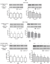
Sperm were incubated at 35°C in the –5mM Ca2+ or +5mM Ca2+ medium with or without CaMKKs inhibitor STO-609 (1; 10; 50 μM) for 5 min and 15 min. Proteins from sperm lysates were analyzed by western-blotting using anti-phospho-Thr172-AMPKα or anti-AMPKα as primary antibody. Bands for phospho-Thr172-AMPKα were detected at 62kDa (top lanes). Total AMPKα (62kDa) was used as loading control (bottom lanes) and the phosphorylated protein AMPKα (Thr172)/total AMPKα ratio is shown at the bottom. A) Effects of different concentrations of STO-609 on the AMPK phosphorylation in sperm incubated for 5 min or 15 min. The control (Ctrl): sperm incubated with or without Ca2+ in the absence of STO-609. B) Effects of 10μM STO-609 on the AMPK phosphorylation in sperm incubated for 5, 15, 30, 60 min. The control (Ctrl): sperm incubated 0 min with or without Ca2+ in the absence of STO-609. Results express the mean ± SEM of the mean from 6 different experiments. Different superscripts indicate statistically significant differences (P < 0.05).
Fig 9
Fig 9. Effect of extracellular Ca2+ on the CaMKI phosphorylation in chicken sperm.
Sperm incubated at 35°C for 5 min in the –5mM Ca2+ or +5mM Ca2+ medium with or without CaMKKs inhibitor STO-609. The control (Ctrl): sperm incubated in the absence both +5mM Ca2+ and 10μM STO-609. Proteins from sperm lysates were analyzed by western-blotting using anti-phospho-Thr177-CaMKI or anti-CaMKI as the primary antibody. Bands for phospho-Thr177-CaMKI were detected at 41kDa (top lanes). Total CaMKI (41kDa) was used as loading control (bottom lanes) and the phosphorylated protein CaMKI (Thr177)/total CaMKI ratio is shown at the bottom. Results express the mean ± SEM of the mean from 4 different experiments. Different superscripts indicate statistically significant differences (P < 0.05).
Fig 10
Fig 10. Effects of STO-609 on the CaMKI phosphorylation in chicken sperm.
Sperm incubated at 35°C in the –5mM Ca2+ or +5mM Ca2+ medium with or without CaMKKs inhibitor STO-609 (0; 1; 10; 50 μM) at 5 min and 15 min. The control (Ctrl): sperm incubated with or without Ca2+ in the absence of STO-609. Proteins from sperm lysates were analyzed by western-blotting using anti-phospho-Thr177-CaMKI or anti-CaMKI as the primary antibody. Bands for phospho-Thr177-CaMKI were detected at 41kDa (top lanes). Total CaMKI (41kDa) was used as loading control (bottom lanes) and the phosphorylated protein CaMKI (Thr177)/total CaMKI ratio is shown at the bottom. Results express the mean ± SEM of the mean from 4 different experiments. Different superscripts indicate statistically significant differences (P<0.05).
Fig 11
Fig 11. Effects of STO-609 on the CaMKIV phosphorylation in chicken sperm.
Sperm incubated at 35°C in the –5mM Ca2+ or +5mM Ca2+ medium with or without CaMKKs inhibitor STO-609 (0; 1; 10; 50 μM) at 5 min and 15 min. The control (Ctrl): sperm incubated with or without Ca2+ in the absence of STO-609. Proteins from sperm lysates were analyzed by western-blotting using anti-phospho-Thr196-CaMKIV or anti-CaMKIV as the primary antibody. Bands for phospho-Thr196-CaMKIV were detected at 60kDa (top lanes). Total CaMKIV (60kDa) was used as loading control (bottom lanes) and the phosphorylated protein CaMKIV (Thr196)/total CaMKIV ratio is shown at the bottom. Results express the mean ± SEM of the mean from 4 different experiments. There were no significant differences between treatments (P<0.05).
Fig 12
Fig 12. Proposed scheme of Ca2+ signaling pathways leading to regulation of AMPK activity in avian sperm.
This schematic diagram shows potential mechanisms of calcium role through CaMKK stimulation in chicken sperm. They all activate AMPK and CaMKI, which leads to metabolic improvements, leading themselves to the control of sperm functions. Solid arrows and blocked arrows illustrate more established relationships between stimuli, signals, improved metabolic status and control of avian sperm functions. Solid black arrows indicate Ca2+/CaMKKs/AMPK signaling pathway. Dashed black arrows indicate Ca2+/CaM/CaMKKs/CaMKI signaling pathway. Curved arrows (black) indicate AMPK phosphorylation, CaMKI phosphorylation; dashed grey arrows with a question mark are used for hypotheses.

Similar articles

Cited by

References

    1. Woods A, Dickerson K, Heath R, Hong SP, Momcilovic M, Johnstone SR, et al. (2005) Ca2+/calmodulin-dependent protein kinase kinase-beta acts upstream of AMP-activated protein kinase in mammalian cells. Cell Metab 2: 21–33. - PubMed
    1. Hawley SA, Pan DA, Mustard KJ, Ross L, Bain J, Edelman AM, et al. (2005) Calmodulin-dependent protein kinase kinase-beta is an alternative upstream kinase for AMP-activated protein kinase. Cell Metab 2: 9–19. - PubMed
    1. Blesbois E, Brillard JP (2007) Specific features of in vivo and in vitro sperm storage in birds. Animal 1: 1472–1481. 10.1017/S175173110700081X - DOI - PubMed
    1. Blesbois E (2012) Biological features of the avian male gamete and their application to biotechnology of conservation. Journal of Poultry Science, 49: 141–149.
    1. Lemoine M, Dupont J, Guillory V, Tesseraud S, Blesbois E (2009) Potential involvement of several signaling pathways in initiation of the chicken acrosome reaction. Biol Reprod 81: 657–665. 10.1095/biolreprod.108.072660 - DOI - PubMed

Publication types

MeSH terms